
















Copyright: ©Author(s) 2026.
World J Gastrointest Oncol. Oct 15, 2026; 18(10): 123542
Published online Oct 15, 2026. doi: 10.4251/wjgo.123542
Published online Oct 15, 2026. doi: 10.4251/wjgo.123542
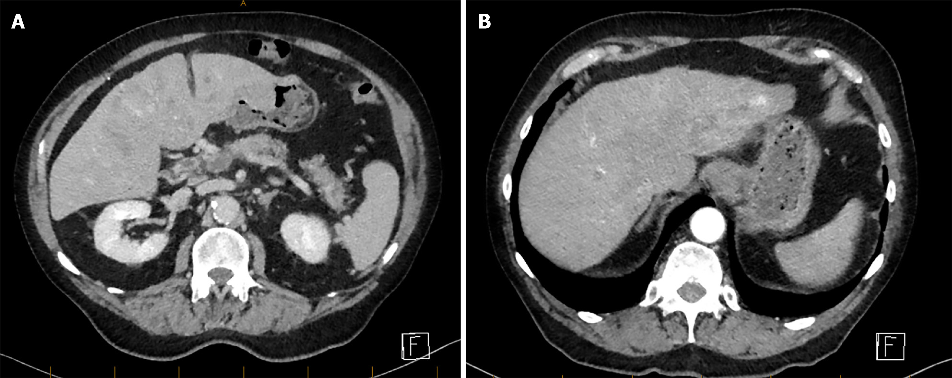
Figure 1 Portal venous phase of computed tomography liver quad phase.
A: Portal vein thrombosis with splenic vein thrombosis; B: Nodularity on the liver surface with segment II liver lesion.
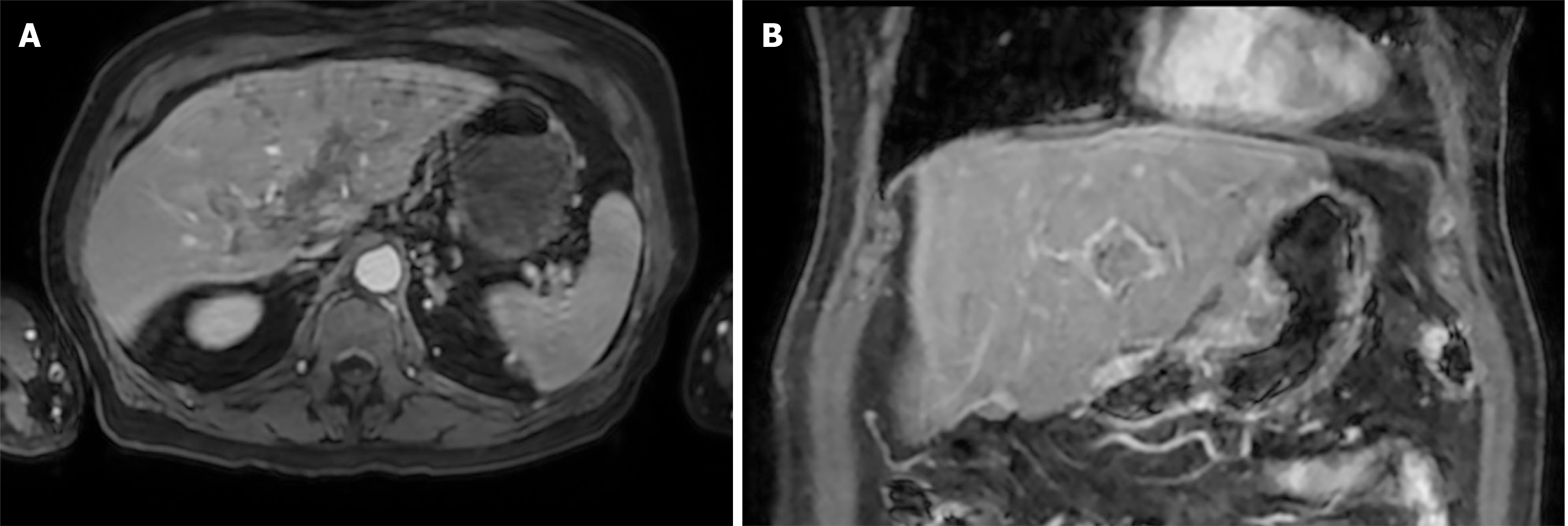
Figure 2 Magnetic resonance imaging liver.
A: Portal vein thrombosis; B: Gastric mass. There is no evidence of hepatocellular carcinoma.
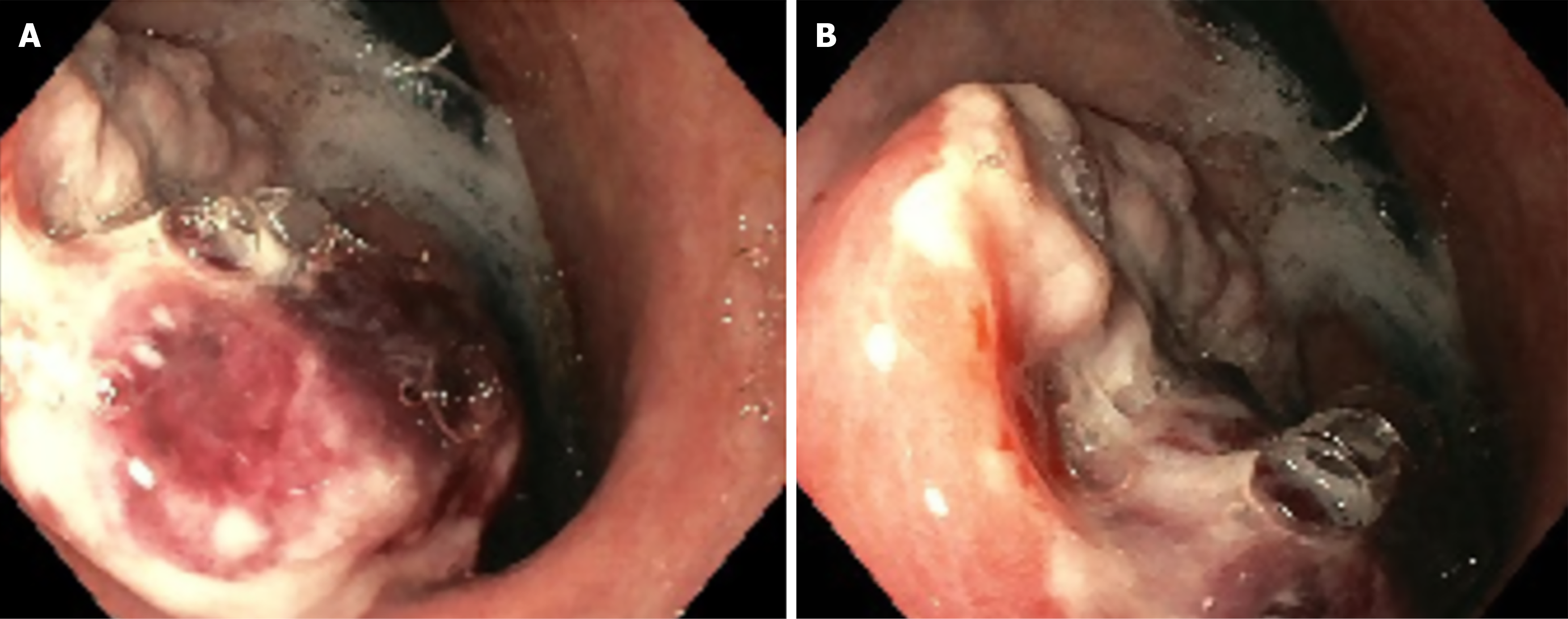
Figure 3 Endoscopic photos of the gastric tumour detected during gastroscopy.
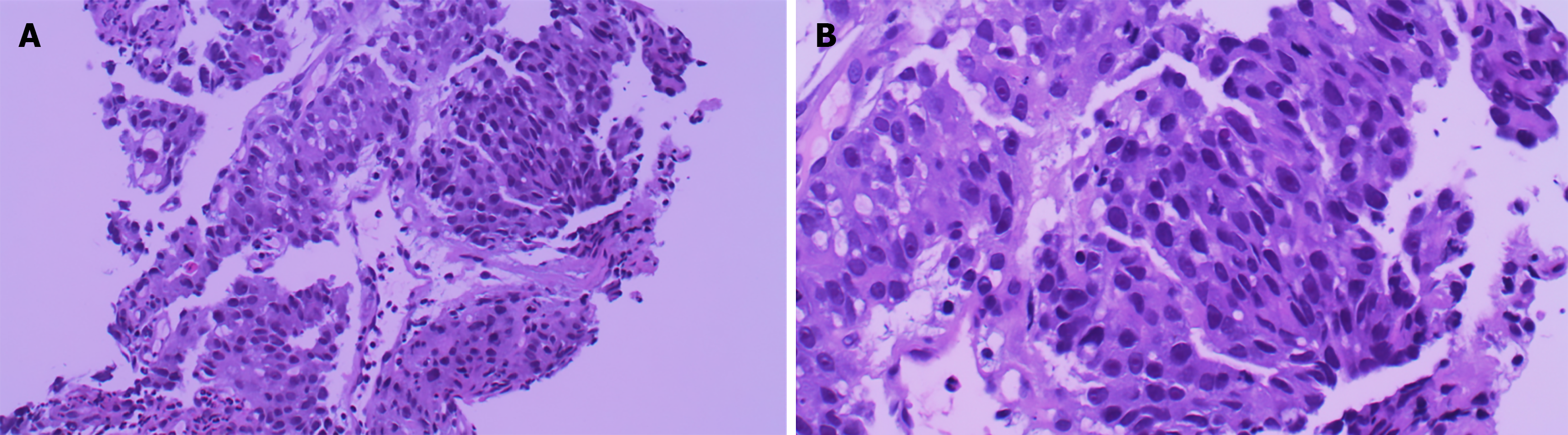
Figure 4 Histopathological findings.
Hematoxylin and eosin staining of gastric biopsy demonstrating hepatoid adenocarcinoma of the gastrointestinal tract. Tumour shows pleomorphic nests and trabecular pattern mimicking that of hepatocellular carcinoma. A: 200 × magnification; B: 400 × magnification.
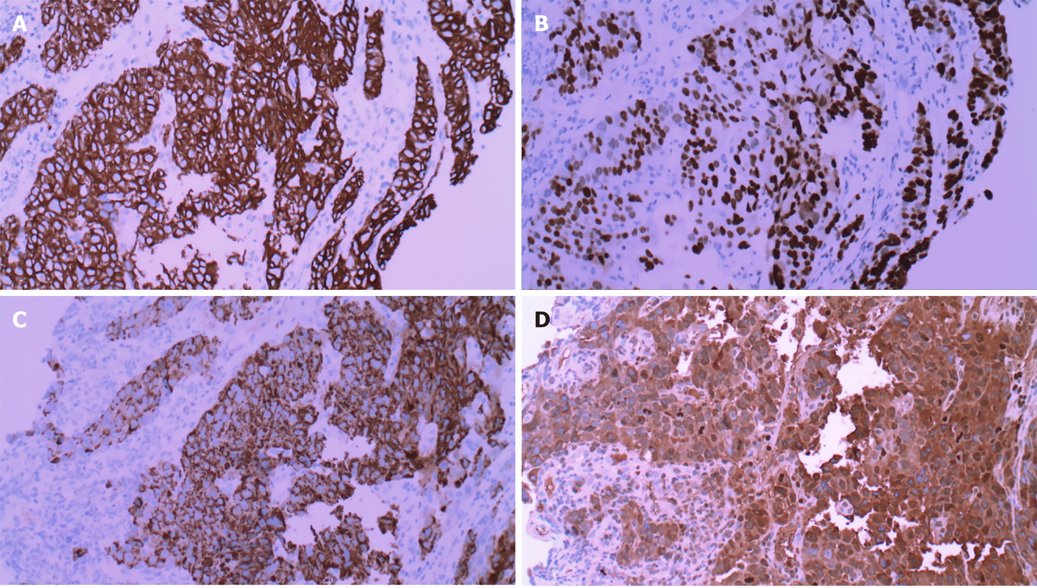
Figure 5 Immunohistochemistry findings.
All images shown at 200 × magnification. A: Cytokeratin AE1AE3 stain strong positive; B: Caudal type homeobox 2 stain positive; C: Hepatocyte paraffin 1 stain positive; D: Alpha-fetoprotein stain positive.
- Citation: Sun SY, Elliott T, Johnstone M, Facey M, Chin JK, Gangadharan SK. Marked elevation of alpha fetoprotein caused by hepatoid adenocarcinoma of stomach: A case report. World J Gastrointest Oncol 2026; 18(10): 123542
- URL: https://www.wjgnet.com/1948-5204/full/v18/i10/123542.htm
- DOI: https://dx.doi.org/10.4251/wjgo.123542