Copyright
©The Author(s) 2025.
World J Gastrointest Oncol. Aug 15, 2025; 17(8): 109424
Published online Aug 15, 2025. doi: 10.4251/wjgo.v17.i8.109424
Published online Aug 15, 2025. doi: 10.4251/wjgo.v17.i8.109424
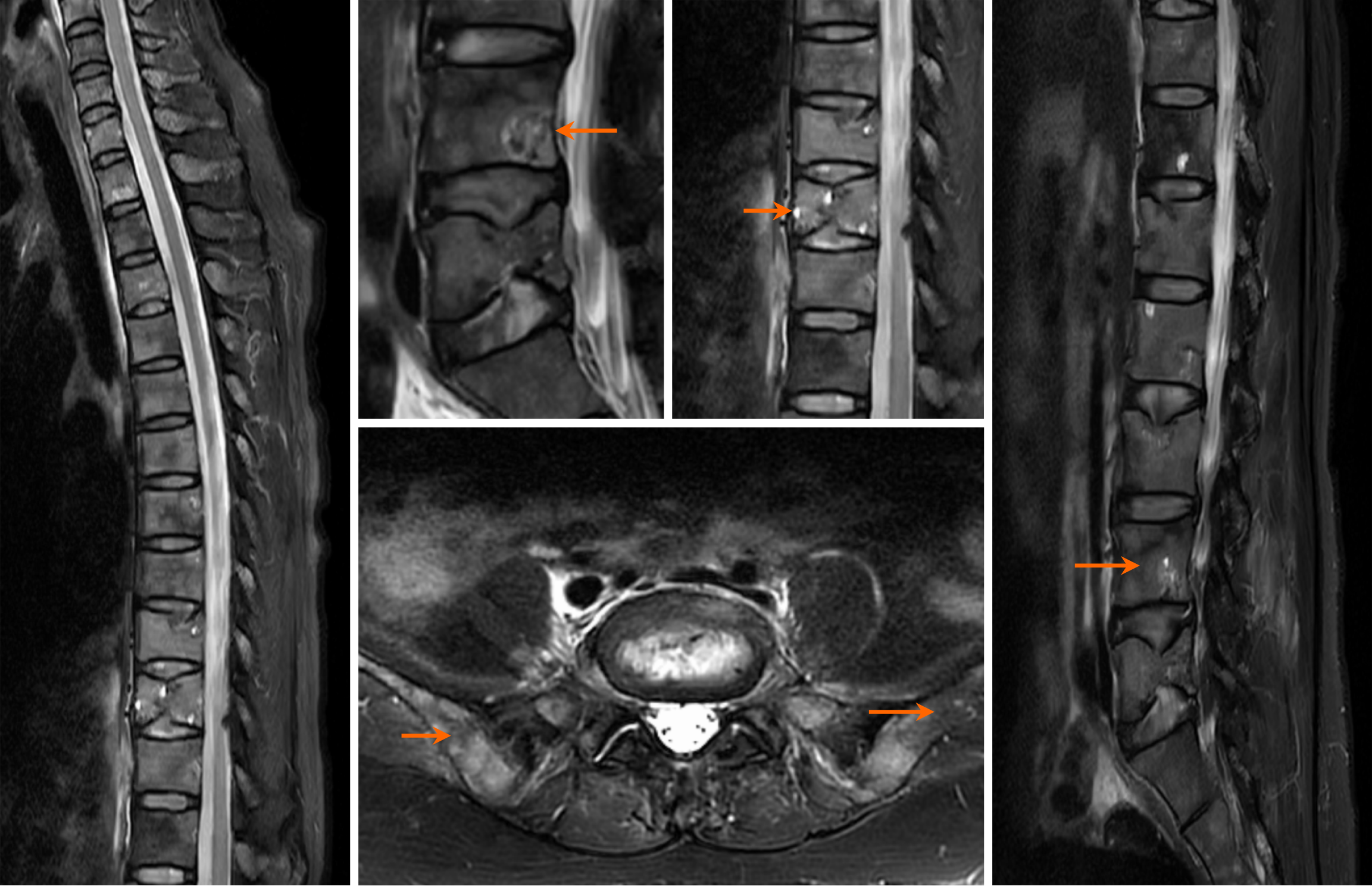
Figure 1
Magnetic resonance imaging showing abnormal signals in the thoracic and lumbar vertebrae and pelvis.
Figure 2 HE staining of pathological sections of gastric biopsy tissue after endoscopy.
A: 100 × magnification; B: 200 × magnification; C: 400 × magnification. Arrows indicate gastric signet ring cells.
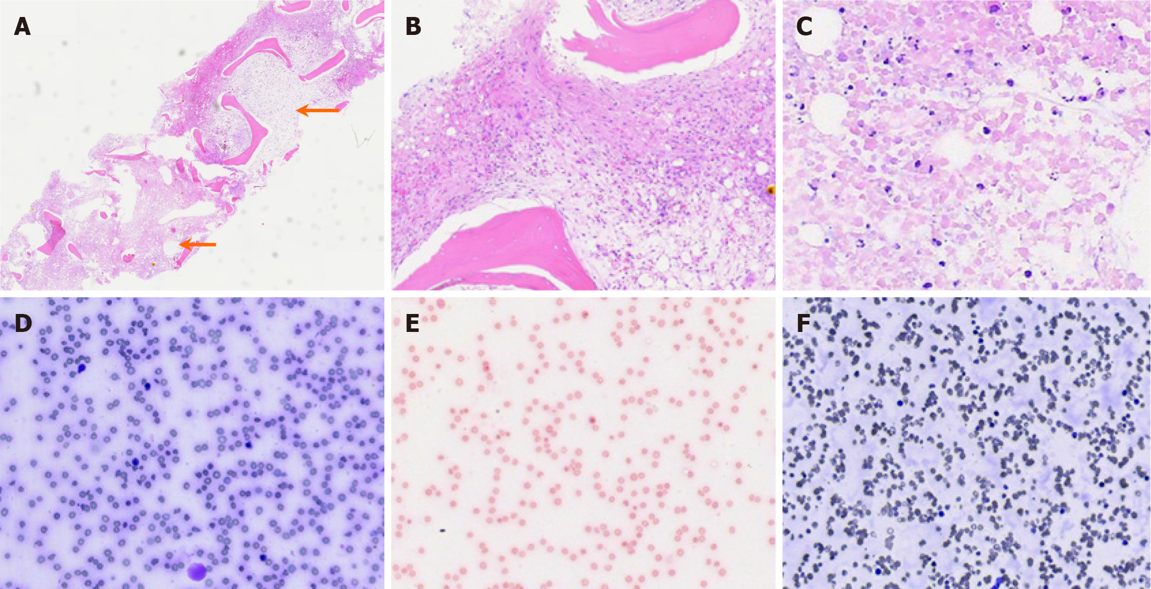
Figure 3 Pathological features of bone marrow failure demonstrating fibrosis, necrosis, dysplastic hematopoiesis, and iron overload.
A: Bone marrow biopsy showing disappearance of typical hematopoietic structure, with necrotic and fibrotic areas (HE staining; 100 ×); B: Enlarged view of fibrotic area showing fibroblast and capillary proliferation with red blood cell extravasation (HE staining; 400 ×); C: Enlarged view of the necrotic area showing extensive coagulative necrosis with some cells exhibiting eccentric nuclei (suspected signet ring cells) and nuclear debris (HE staining; 800 ×); D: Bone marrow smear showing low-level hyperplasia, decreased granulocytic series, active erythroid series, absence of megakaryocytes, and bone marrow particles (Wright staining; 400 ×); E: Bone marrow smear showing absence of bone marrow particles and 74% positive iron staining (Iron staining; 400 ×); F: Peripheral blood smear showing normal proportions of mature granulocytes and lymphocytes, with visible immature red and white blood cells and unevenly distributed platelets (Wright staining; 400 ×).
- Citation: Sun W, Chen XC, Wang H, Chang WY, He Y, Lin ZH, Jia H, Zhang XM, Liu H. Bone marrow metastasis of gastric signet ring cell carcinoma complicated by thrombotic microangiopathy: A case report. World J Gastrointest Oncol 2025; 17(8): 109424
- URL: https://www.wjgnet.com/1948-5204/full/v17/i8/109424.htm
- DOI: https://dx.doi.org/10.4251/wjgo.v17.i8.109424


