Copyright
©The Author(s) 2025.
World J Gastrointest Oncol. Jun 15, 2025; 17(6): 107568
Published online Jun 15, 2025. doi: 10.4251/wjgo.v17.i6.107568
Published online Jun 15, 2025. doi: 10.4251/wjgo.v17.i6.107568
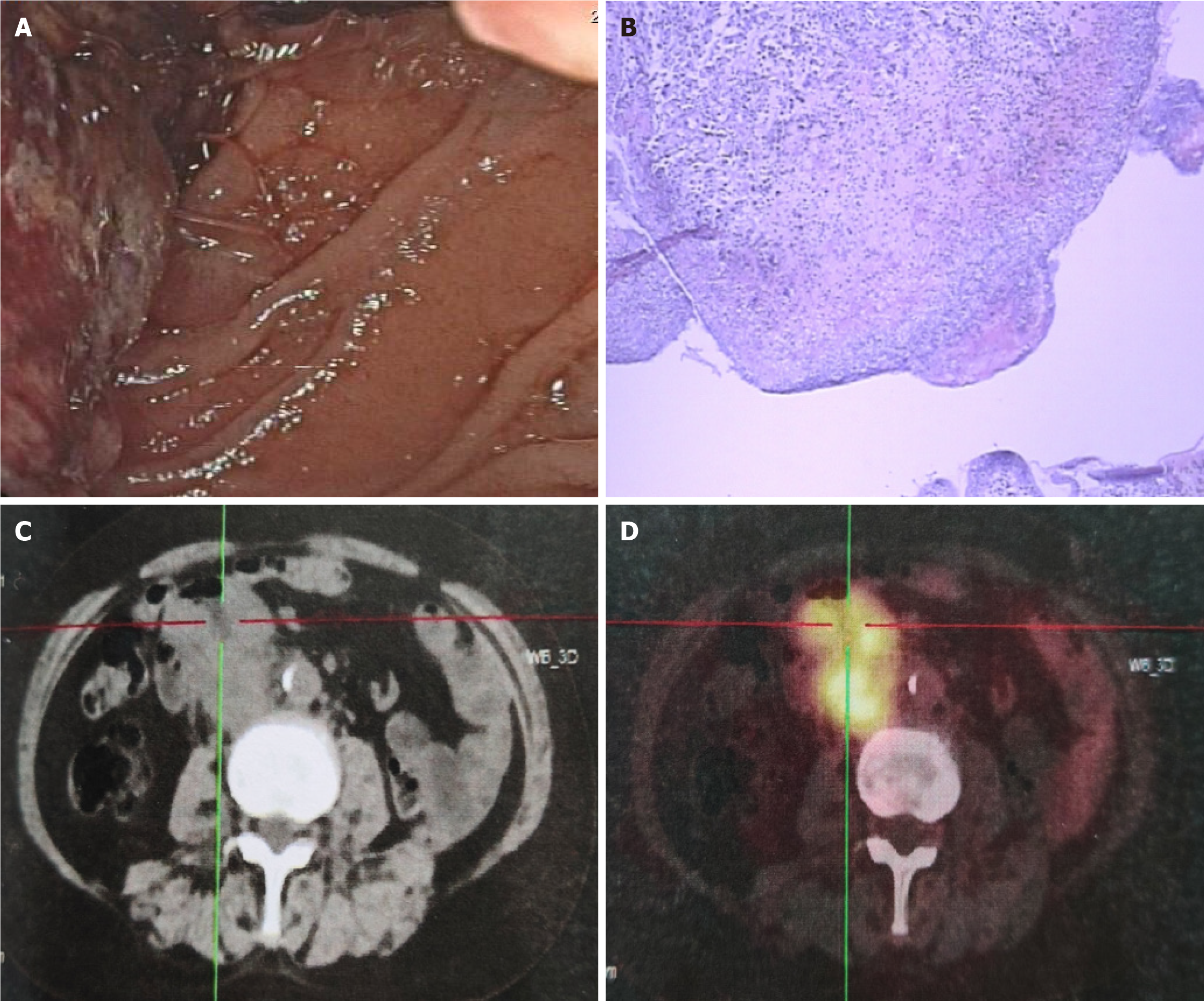
Figure 1 Before receiving chemotherapy.
A: Upper gastrointestinal endoscopy showed a mass in the descending duodenum with bleeding; B: Hematoxylin-eosin staining showed adenocarcinoma; C and D: Positron emission tomography/computed tomography examination showed uptake of 18F-fluorodeoxyglucose in the descending and horizontal segments of the duodenum, around the intestinal wall, and retroperitoneum.
Figure 2 After receiving chemotherapy, immediately before pembrolizumab.
A: Upper gastrointestinal endoscopy showed a mass in the descending duodenum with numerous violaceous necrotic tissue and blood clots on the surface; B: Computed tomography (CT) showed a duodenal mass with possible retroperitoneal lymph node metastasis; C: CT showed dilated right renal pelvis and abdominal ureter.
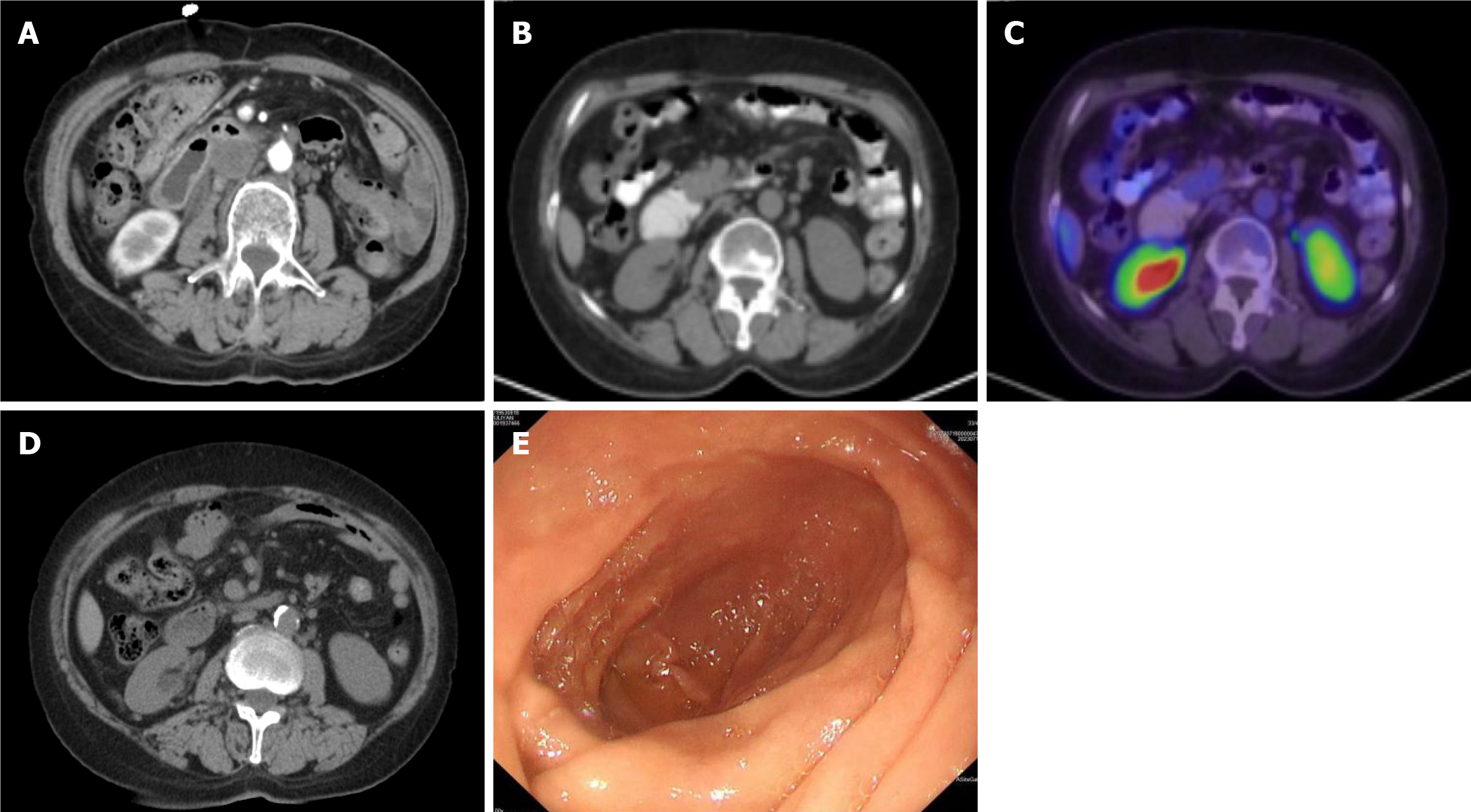
Figure 3 After receiving pembrolizumab.
A: After receiving 2 cycles of pembrolizumab. computed tomography (CT) showed that the duodenal mass was significantly smaller than before; B and C: After receiving pembrolizumab for 2 years. Positron emission tomography/CT showed low density at the lateral pancreatic head attached to the duodenum and no increase in metabolism; D and E: After receiving 4 years of pembrolizumab. CT revealed no thickening of the duodenal wall (D). Upper gastrointestinal endoscopy showed smooth mucosa in the descending duodenum (E).
- Citation: Kong XL, Lu XW, Dong SQ, Liu J, Zheng L, Chen LL, An ZH, Gao LM, Cao JL. Descending duodenal adenocarcinoma treated with pembrolizumab resulting in complete clinical response: A case report and literature review. World J Gastrointest Oncol 2025; 17(6): 107568
- URL: https://www.wjgnet.com/1948-5204/full/v17/i6/107568.htm
- DOI: https://dx.doi.org/10.4251/wjgo.v17.i6.107568


