Copyright
©The Author(s) 2023.
World J Gastrointest Oncol. Aug 15, 2023; 15(8): 1497-1504
Published online Aug 15, 2023. doi: 10.4251/wjgo.v15.i8.1497
Published online Aug 15, 2023. doi: 10.4251/wjgo.v15.i8.1497
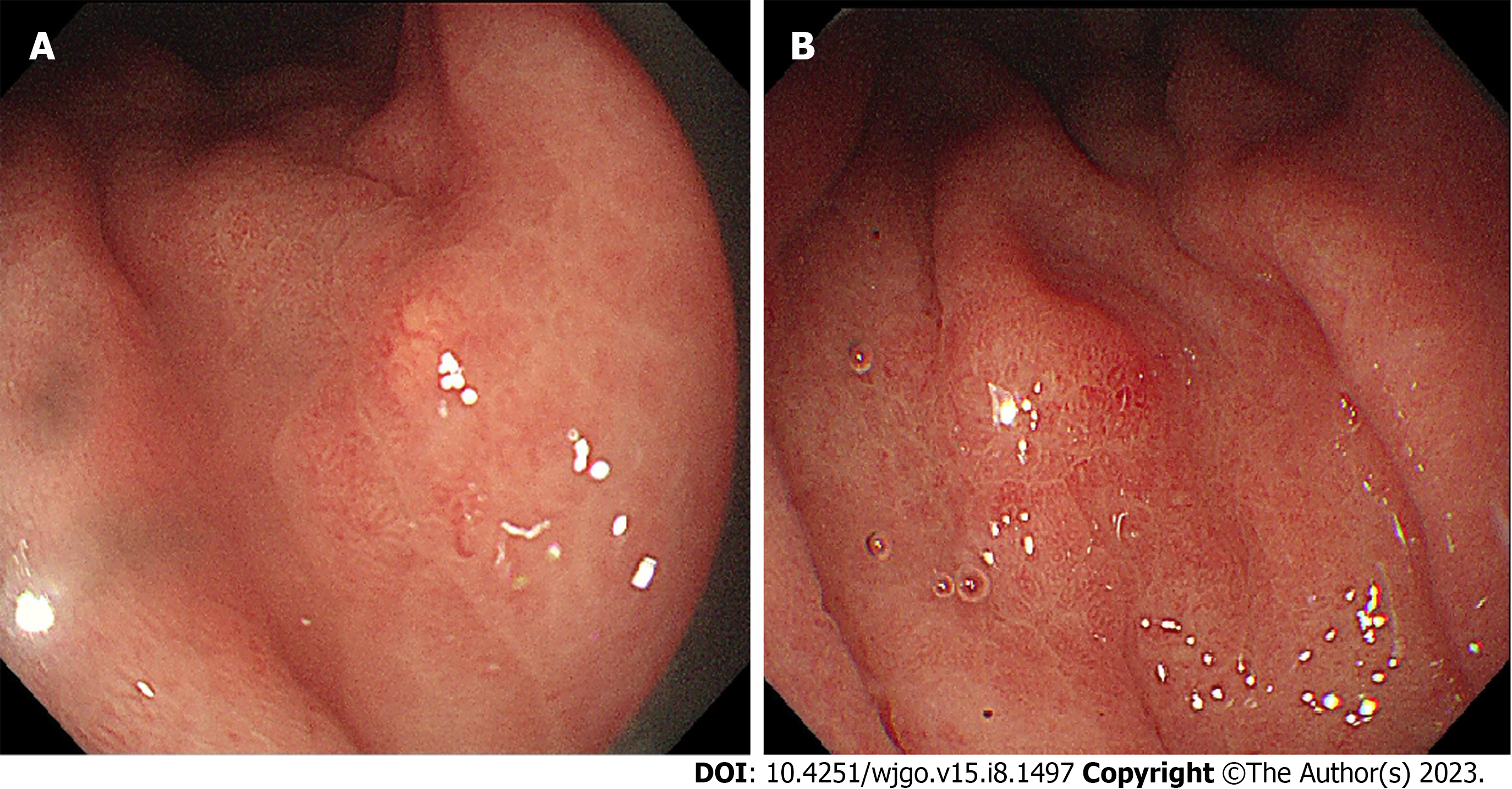
Figure 1 Polyps and a ulcer of the gastric mucosa could be seen under an endoscope.
A: Gastric mucosa atrophy and polyps were seen; B: A superficial ulcer with central scar-like changes was seen in the large curvature of the central part of the stomach.
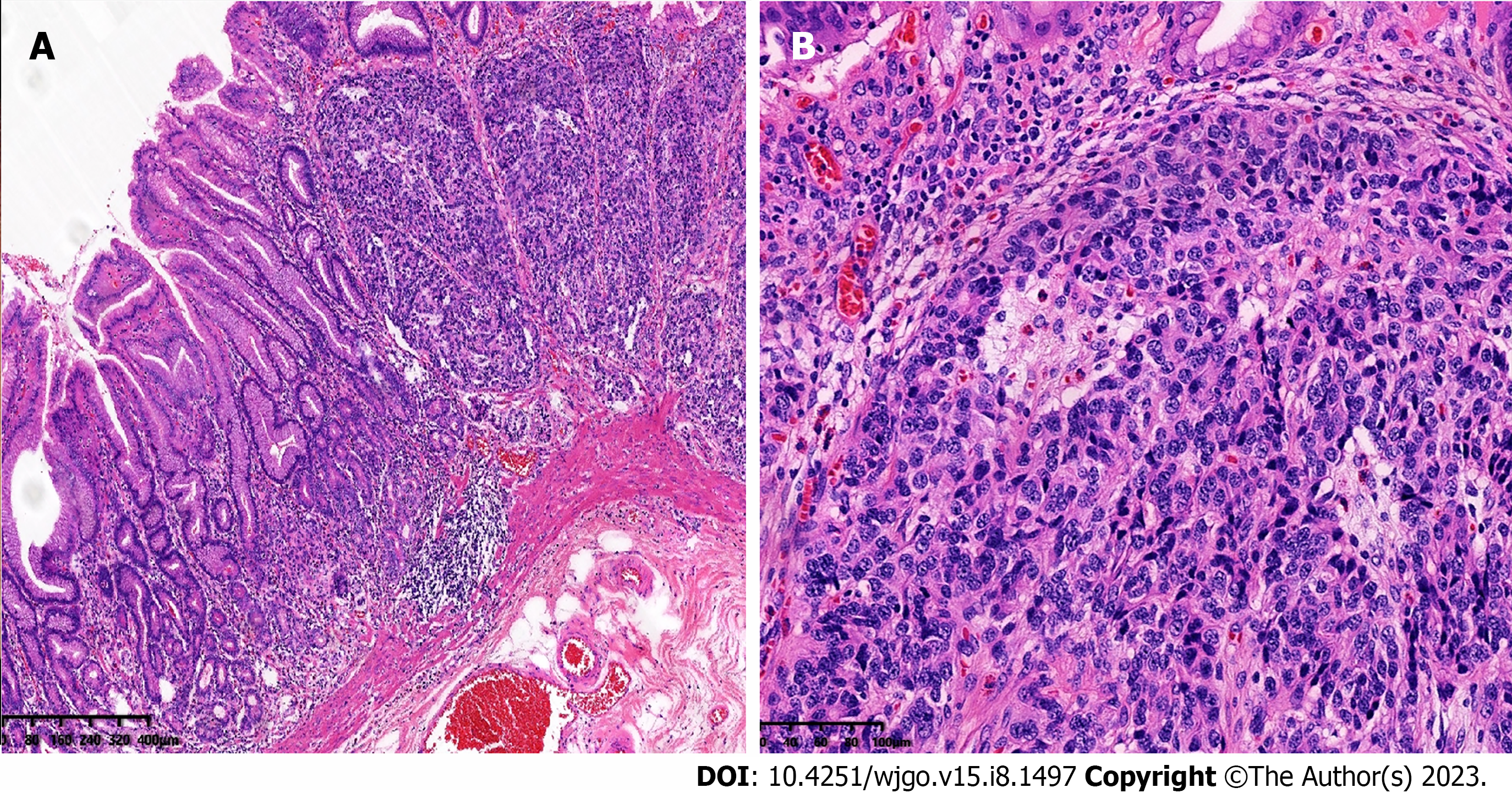
Figure 2 Pathological morphology of gastric neuroendocrine tumors.
A: Under low-power magnification, a neuroendocrine tumor was shown to infiltrate into the surrounding tissues; B: Under high-power magnification, tumor cells were rich in blood sinuses.
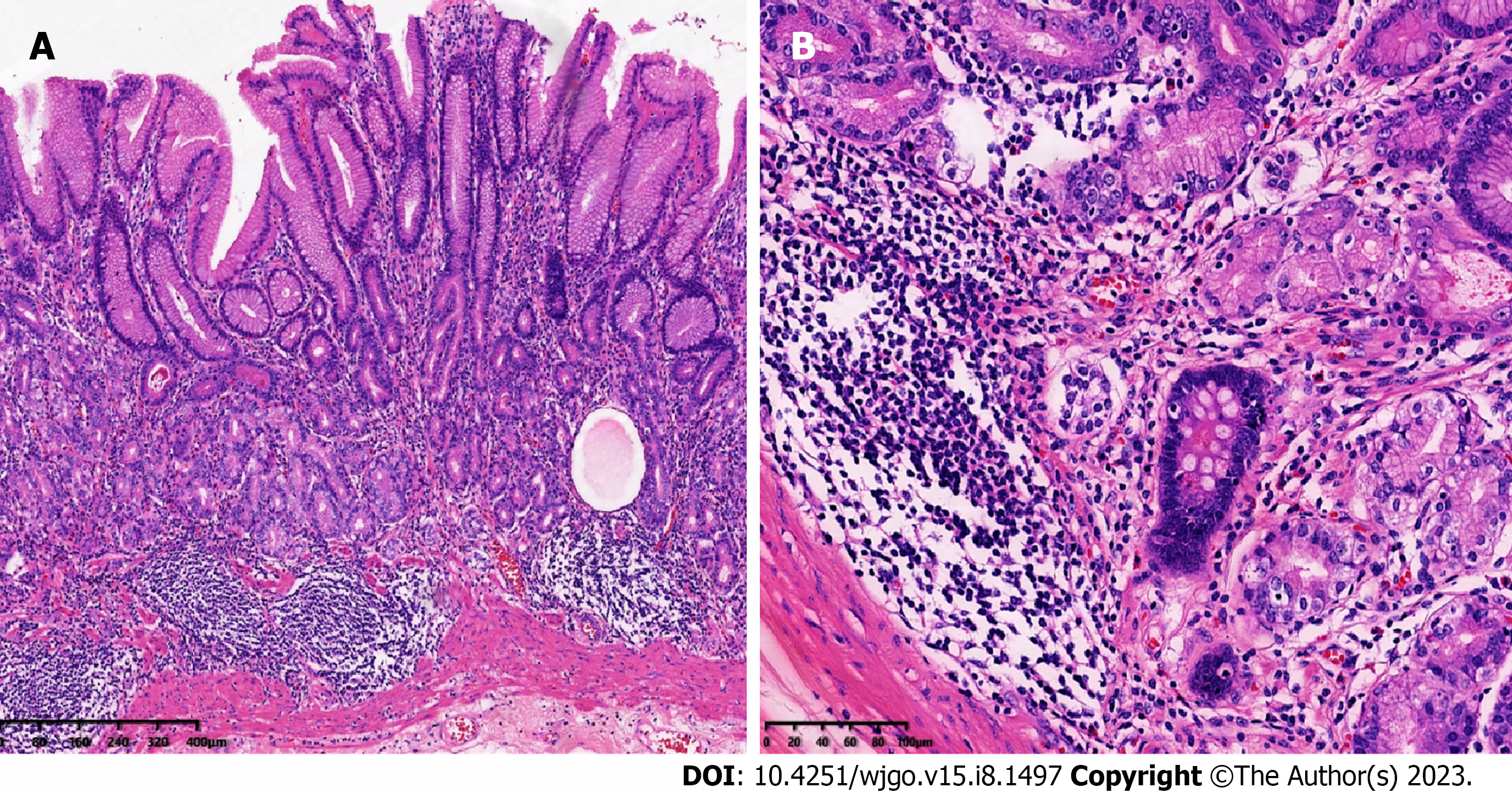
Figure 3 Pathological changes of atrophic gastritis in the gastric corpus mucosa.
A: Under low-power magnification, a reduction in the number of gastric fundus glands was seen; B: Under high-power magnification, gland intestinal metaplasia and lymphocyte infiltration were seen.
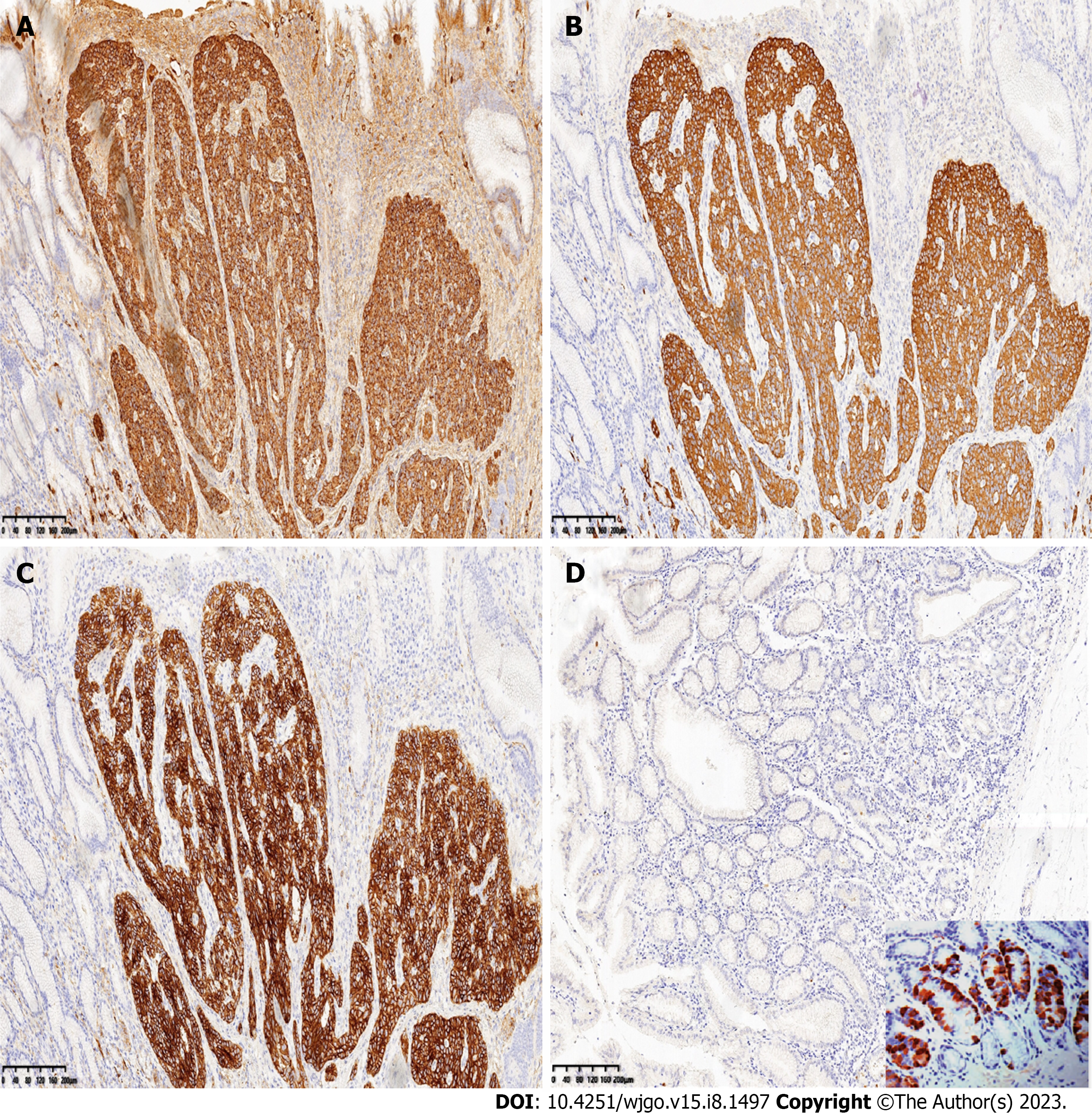
Figure 4 Immunohistochemical findings.
A-C: The tumor cells were strongly positive for CgA (A), Syn (B), and CD56 (C); D: Immunohistochemical staining showed that gastrin was absent. An image showing positive immunohistochemical staining for gastrin is presented in the lower right corner as a positive control for comparison.
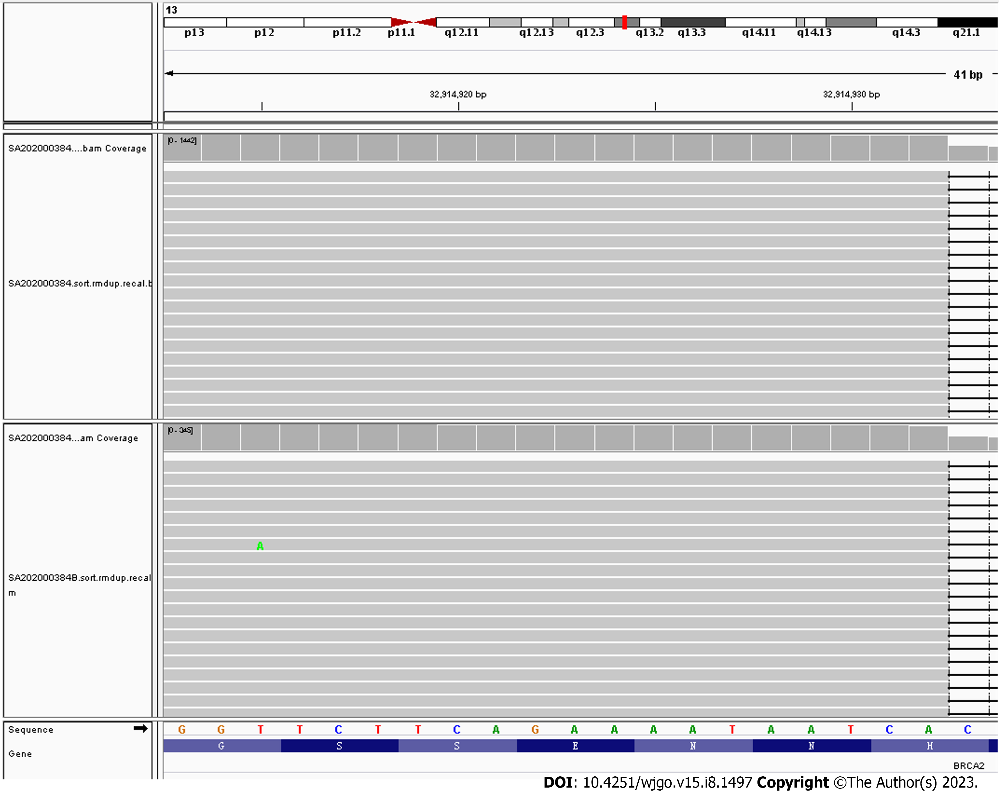
Figure 5 Next-generation sequencing showed the presence of a heterozygous germline mutation in BRCA2 exon 11, c.
6443_644del, (p_s2148Yfs*2). The upper and lower parts in the figure are tumor and control, respectively.
- Citation: Zhang HF, Zheng Y, Wen X, Zhao J, Li J. Gastric neuroendocrine tumors in a BRCA2 germline mutation carrier: A case report. World J Gastrointest Oncol 2023; 15(8): 1497-1504
- URL: https://www.wjgnet.com/1948-5204/full/v15/i8/1497.htm
- DOI: https://dx.doi.org/10.4251/wjgo.v15.i8.1497


