Copyright
©The Author(s) 2023.
World J Gastrointest Oncol. Jul 15, 2023; 15(7): 1311-1316
Published online Jul 15, 2023. doi: 10.4251/wjgo.v15.i7.1311
Published online Jul 15, 2023. doi: 10.4251/wjgo.v15.i7.1311
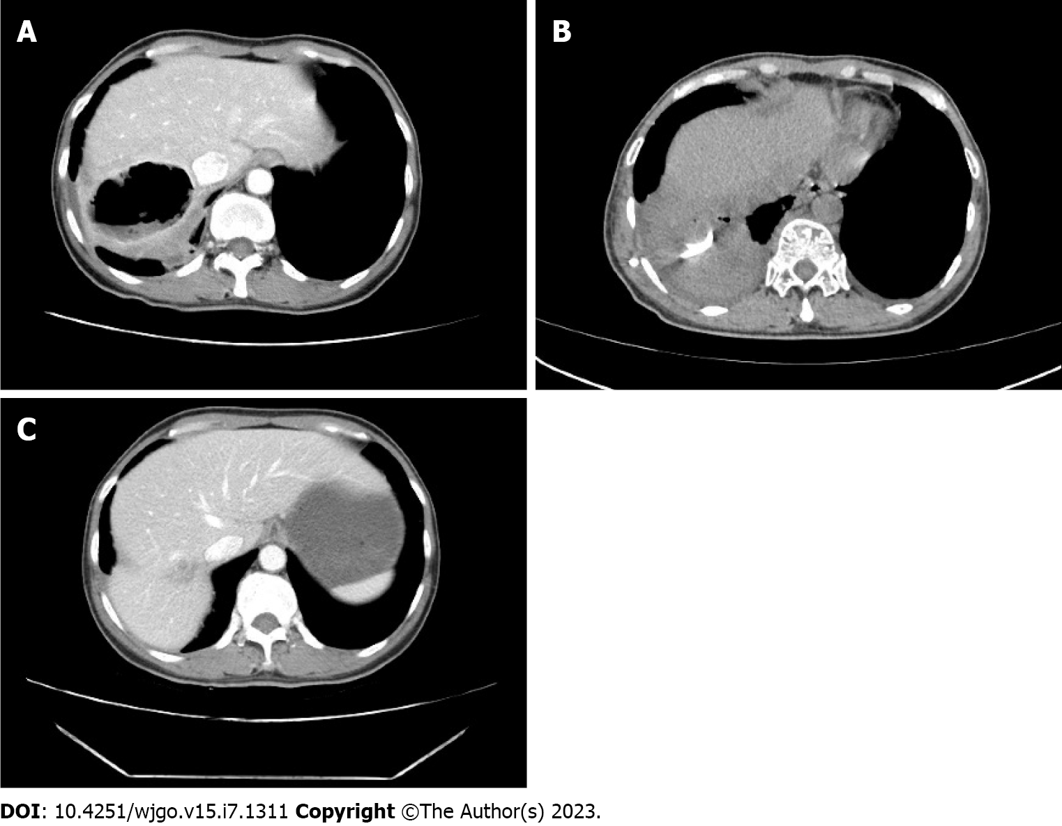
Figure 1 Contrast-enhanced abdominal computed tomography.
A: Contrast-enhanced abdominal computed tomography (CT) on December 19, 2022 showed liver abscess, with a maximum section of 6.9 cm × 6.0 cm, accompanied by fluid and gas; B: Contrast-enhanced abdominal CT on December 29, 2022 showed a cavity shadow in the right posterior lobe of the liver, and a drainage tube shadow was seen inside the cavity. The cavity was smaller than before, and no gas-liquid level was observed; C: Contrast-enhanced abdominal CT on March 27, 2023 showed the filling of solid components in the primary cavity of the right posterior lobe of the liver, and the enhancement of the cavity wall was relatively uniform.
- Citation: Hu W, Lin X, Qian M, Du TM, Lan X. Treatment of Candida albicans liver abscess complicated with COVID-19 after liver metastasis ablation: A case report. World J Gastrointest Oncol 2023; 15(7): 1311-1316
- URL: https://www.wjgnet.com/1948-5204/full/v15/i7/1311.htm
- DOI: https://dx.doi.org/10.4251/wjgo.v15.i7.1311


