Copyright
©The Author(s) 2023.
World J Gastrointest Oncol. Nov 15, 2023; 15(11): 1998-2016
Published online Nov 15, 2023. doi: 10.4251/wjgo.v15.i11.1998
Published online Nov 15, 2023. doi: 10.4251/wjgo.v15.i11.1998
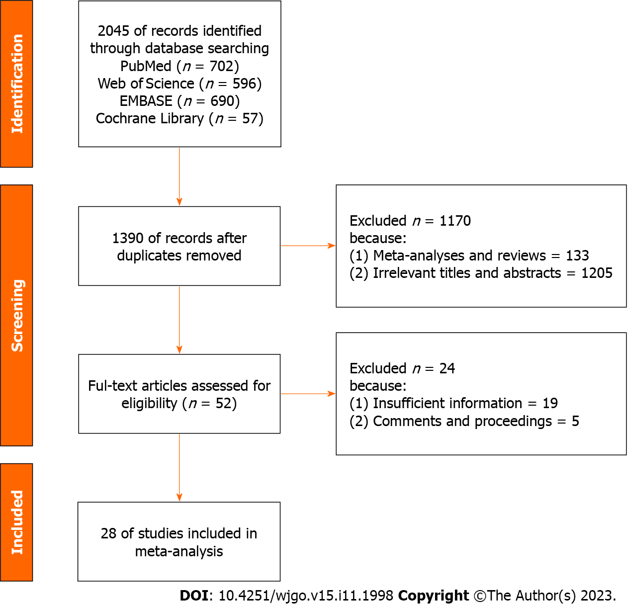
Figure 1 Flowchart of the search process.
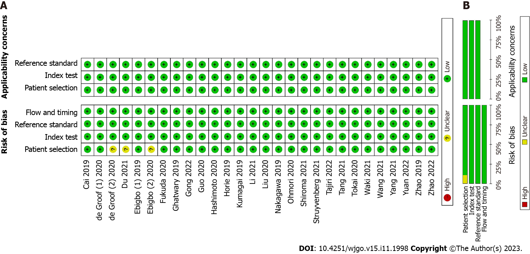
Figure 2 Methodological quality assessment.
A: Summary graph of quality in the methodology; B: Summary table of quality in the methodology.
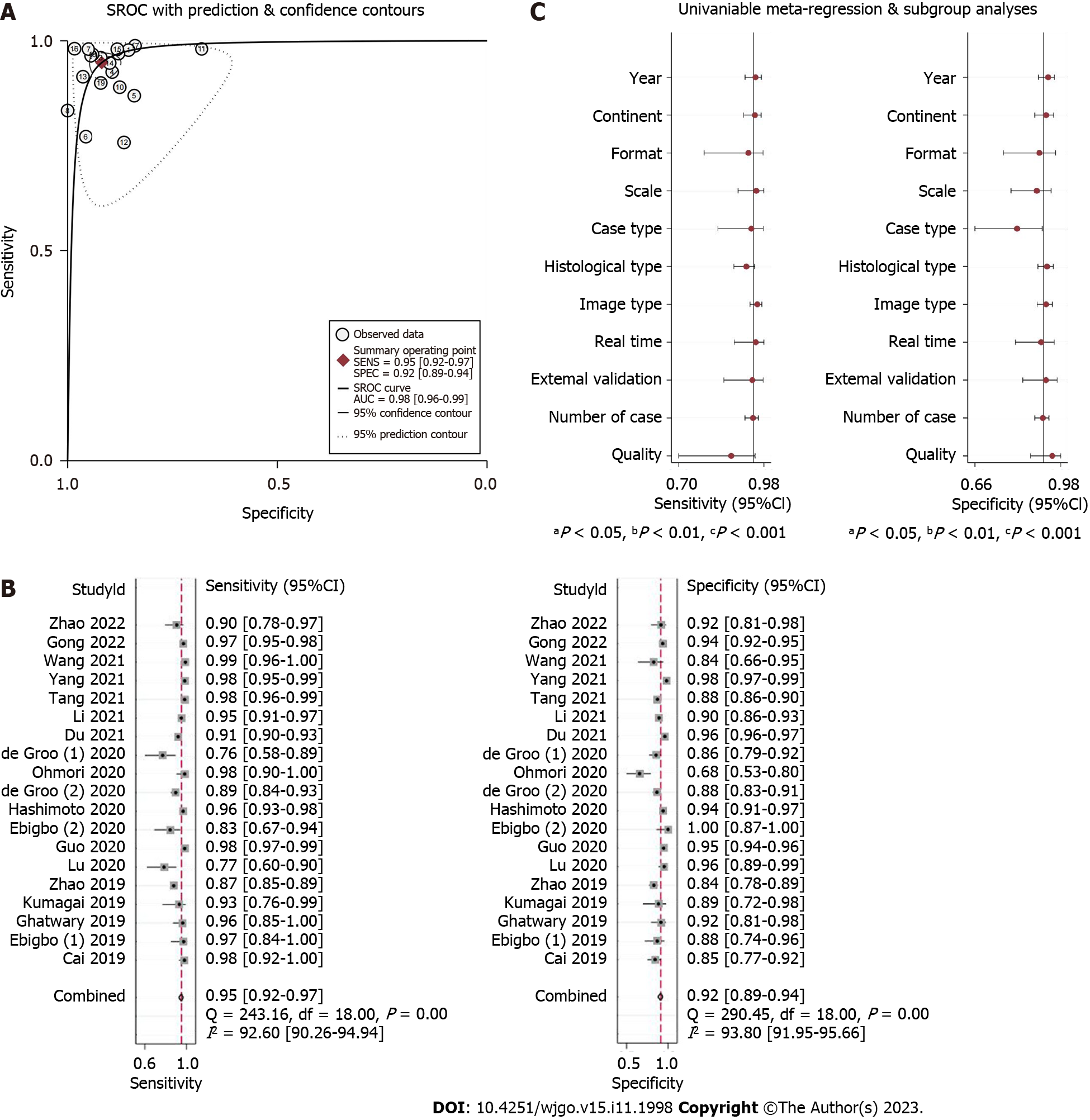
Figure 3 Summary of the receiver operating characteristic, forest plots, and univariable meta-regression plot of convolutional neural network for the diagnosis of esophageal cancer or high-grade dysplasia based on still images.
A: Summary of the receiver operating characteristic of convolutional neural network (CNN) for the diagnosis of esophageal cancer or high-grade dysplasia (HGD) based on still images; B: Coupled forest plots for the sensitivity and specificity of CNN in the diagnosis of esophageal cancer or HGD based on still images; C: Univariable meta-regression plot of CNN for the diagnosis of esophageal cancer or HGD based on still images. CI: Confidence interval; SROC: Summary receiver operating characteristic.
Figure 4 Forest plots of convolutional neural network and endoscopist results for the diagnosis of esophageal cancer or high-grade dysplasia based on still images.
A: Forest plot of the sensitivity by endoscopists for the diagnosis of esophageal cancer or high-grade dysplasia (HGD) based on still images; B: Forest plot of the specificity by endoscopists for the diagnosis of esophageal cancer or HGD based on still images; C: Forest plot of the sensitivity by convolutional neural network (CNN) for the diagnosis of esophageal cancer or HGD based on still images; D: Forest plot of the specificity by CNN for the diagnosis of esophageal cancer or HGD based on still images. CI: Confidence interval.
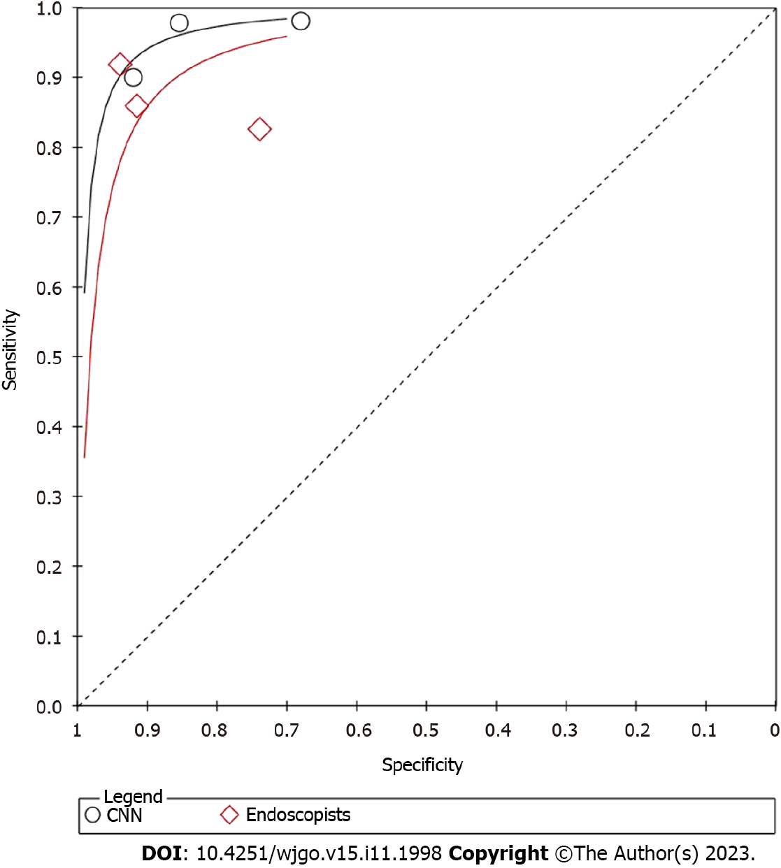
Figure 5 Summary of the receiver operating characteristic by convolutional neural network and endoscopists for the diagnosis of esophageal cancer or high-grade dysplasia based on still images.
CNN: Convolutional neural network.
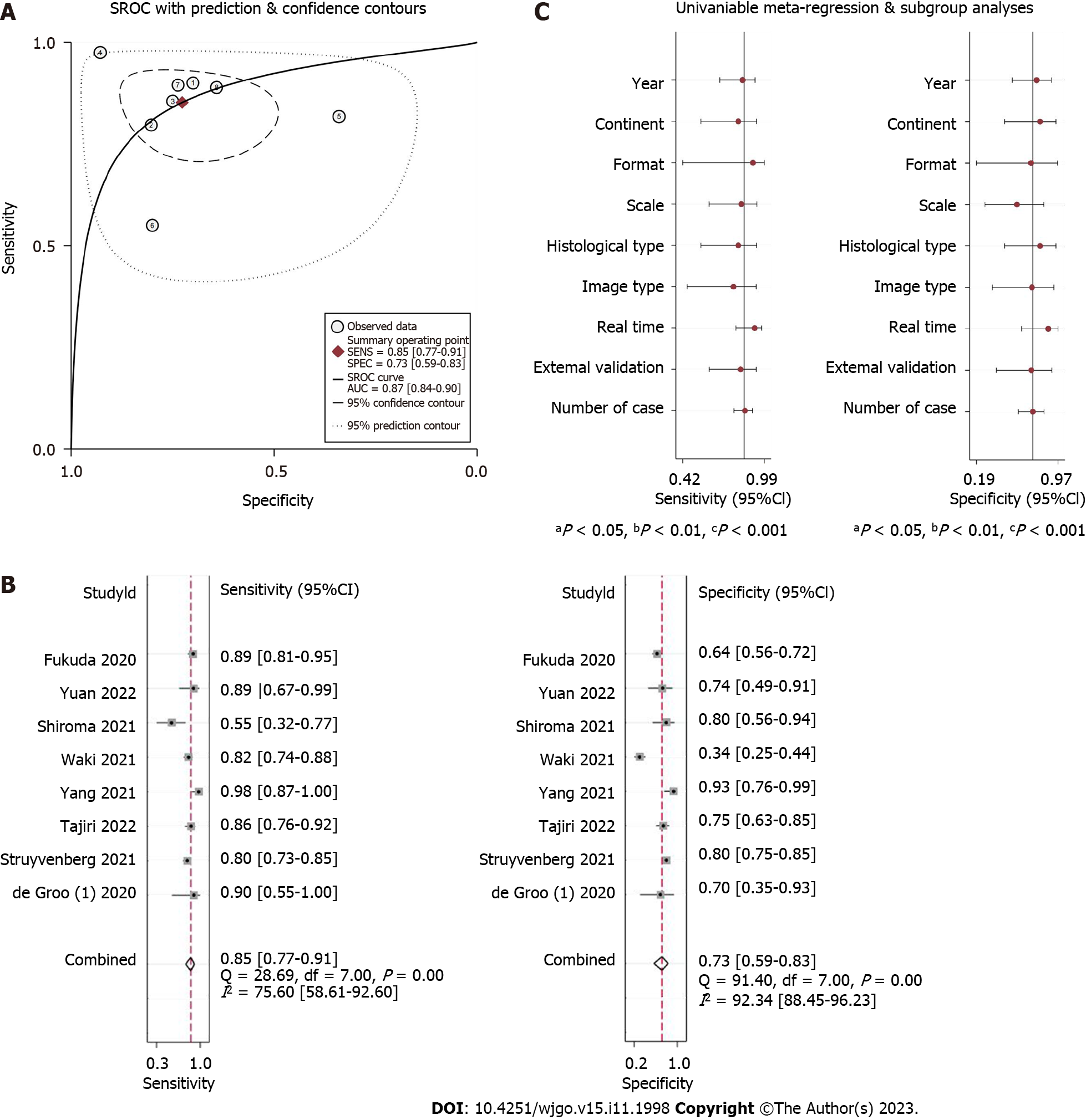
Figure 6 Summary of receiver operating characteristic, forest plots, and univariable meta-regression plot of convolutional neural network in the diagnosis of esophageal cancer or high-grade dysplasia based on videos.
A: Summary of the receiver operating characteristic of convolutional neural network (CNN) for the diagnosis of esophageal cancer or high-grade dysplasia (HGD) based on videos; B: Coupled forest plots of sensitivity and specificity of CNN for the diagnosis of esophageal cancer or HGD based on videos; C: Univariable meta-regression plot of CNN for the diagnosis of esophageal cancer or HGD based on videos. CI: Confidence interval; SROC: Summary receiver operating characteristic.
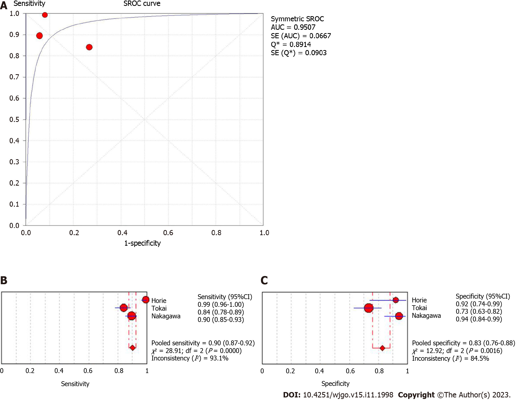
Figure 7 Summary of receiver operating characteristic and forest plots for convolutional neural network in predicting the invasion depth of esophageal cancer.
A: Summary of receiver operating characteristic for convolutional neural network (CNN) in predicting the invasion depth of esophageal cancer; B: Forest plots of sensitivity for CNN in predicting the invasion depth of esophageal cancer; C: Forest plots of specificity for CNN in predicting the invasion depth of esophageal cancer. AUC: Area under the curve; SROC: Summary receiver operating characteristic; CI: Confidence interval.
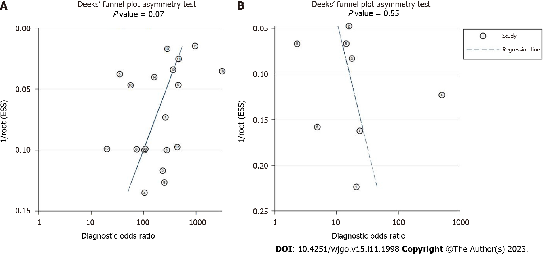
Figure 8 Deeks’ plot of publication bias.
A Deek’s funnel plot of convolutional neural network (CNN) for the diagnosis of esophageal cancer or high-grade dysplasia (HGD) based on still images; B: Deek’s funnel plot of CNN for the diagnosis of esophageal cancer or HGD based on videos.
- Citation: Zhang JQ, Mi JJ, Wang R. Application of convolutional neural network-based endoscopic imaging in esophageal cancer or high-grade dysplasia: A systematic review and meta-analysis. World J Gastrointest Oncol 2023; 15(11): 1998-2016
- URL: https://www.wjgnet.com/1948-5204/full/v15/i11/1998.htm
- DOI: https://dx.doi.org/10.4251/wjgo.v15.i11.1998


