Copyright
©The Author(s) 2023.
World J Gastrointest Oncol. Oct 15, 2023; 15(10): 1807-1822
Published online Oct 15, 2023. doi: 10.4251/wjgo.v15.i10.1807
Published online Oct 15, 2023. doi: 10.4251/wjgo.v15.i10.1807
Figure 1 Gastroscopic and imaging features of gastric cancer concomitant with gastrointestinal stromal tumor.
A: Ulcerative gastric adenocarcinoma (patient 6); B: Pyloric adenocarcinoma (thin arrow) and intraluminal gastrointestinal stromal tumor (GIST; thick arrow) leading to pyloric obstruction (patient 1); C: Giant GIST (9.5 cm × 10.9 cm × 11.7 cm, arrow) showing intraluminal and extraluminal growth, compressing the spleen and left kidney (patient 11); D: Suspicious GIST under gastroscopy (patient 6); E: Suspicious GIST on ultrasound Endoscopy (patient 6).
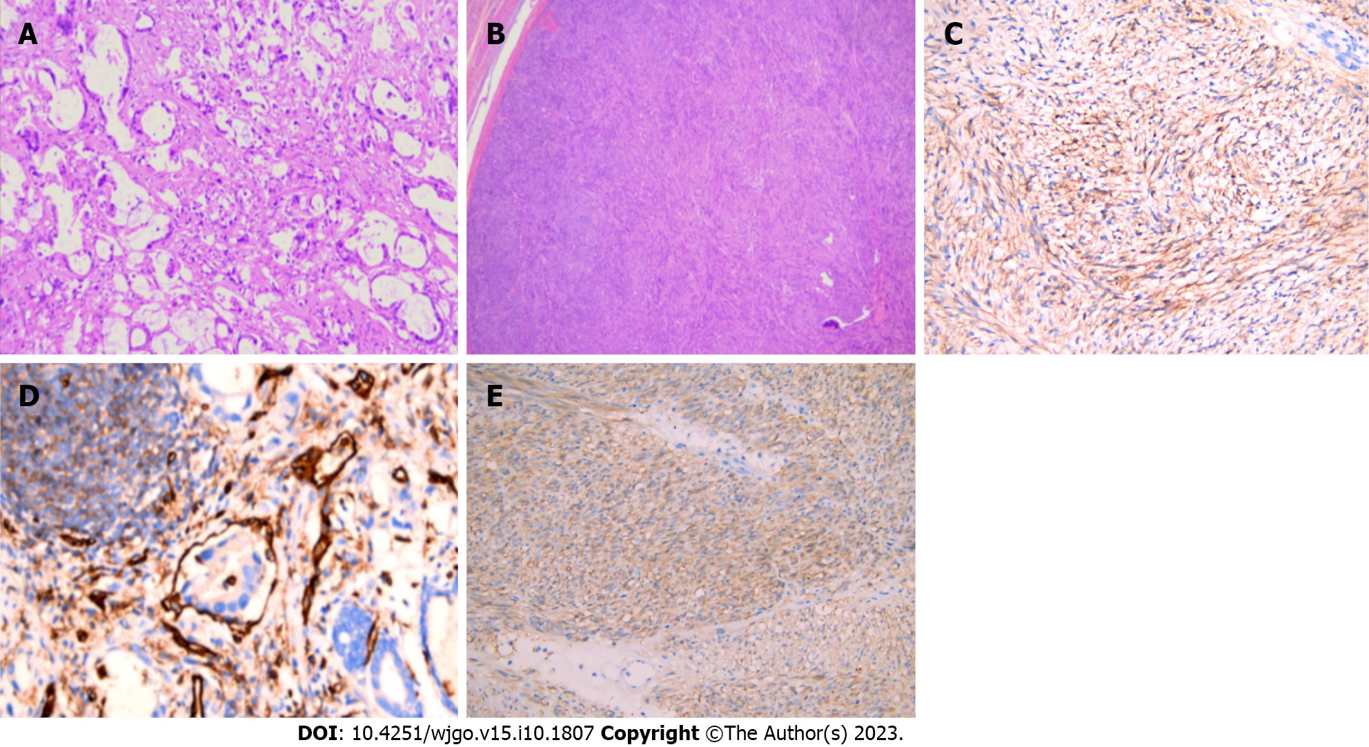
Figure 2 Pathological and immunohistochemical features of gastric cancer concomitant with gastrointestinal stromal tumor.
A: Microscopically showing gastric adenocarcinoma (patient 6; HE, 100 ×); B: Microscopically showing gastric stromal tumor (patient 6; HE, 100 ×); C: CD117 positive under microscope (patient 6; immunohistochemical staining, 200 ×); D: CD34 positive under microscope (patient 6; immunohistochemical staining, 200 ×); E: Dog-1 positive under microscope (patient 6; immunohistochemical staining, 200 ×).
Figure 3 Kaplan-Meier survival curves.
A: 19 patients with gastric cancer (GC) accompanying gastrointestinal stromal tumor (GIST) in this study; B: 46 patients with GC accompanying GIST in this study and the literature reviewed.
- Citation: Liu J, Huang BJ, Ding FF, Tang FT, Li YM. Synchronous occurrence of gastric cancer and gastrointestinal stromal tumor: A case report and review of the literature. World J Gastrointest Oncol 2023; 15(10): 1807-1822
- URL: https://www.wjgnet.com/1948-5204/full/v15/i10/1807.htm
- DOI: https://dx.doi.org/10.4251/wjgo.v15.i10.1807


