Copyright
©The Author(s) 2021.
World J Gastrointest Oncol. Sep 15, 2021; 13(9): 1043-1061
Published online Sep 15, 2021. doi: 10.4251/wjgo.v13.i9.1043
Published online Sep 15, 2021. doi: 10.4251/wjgo.v13.i9.1043
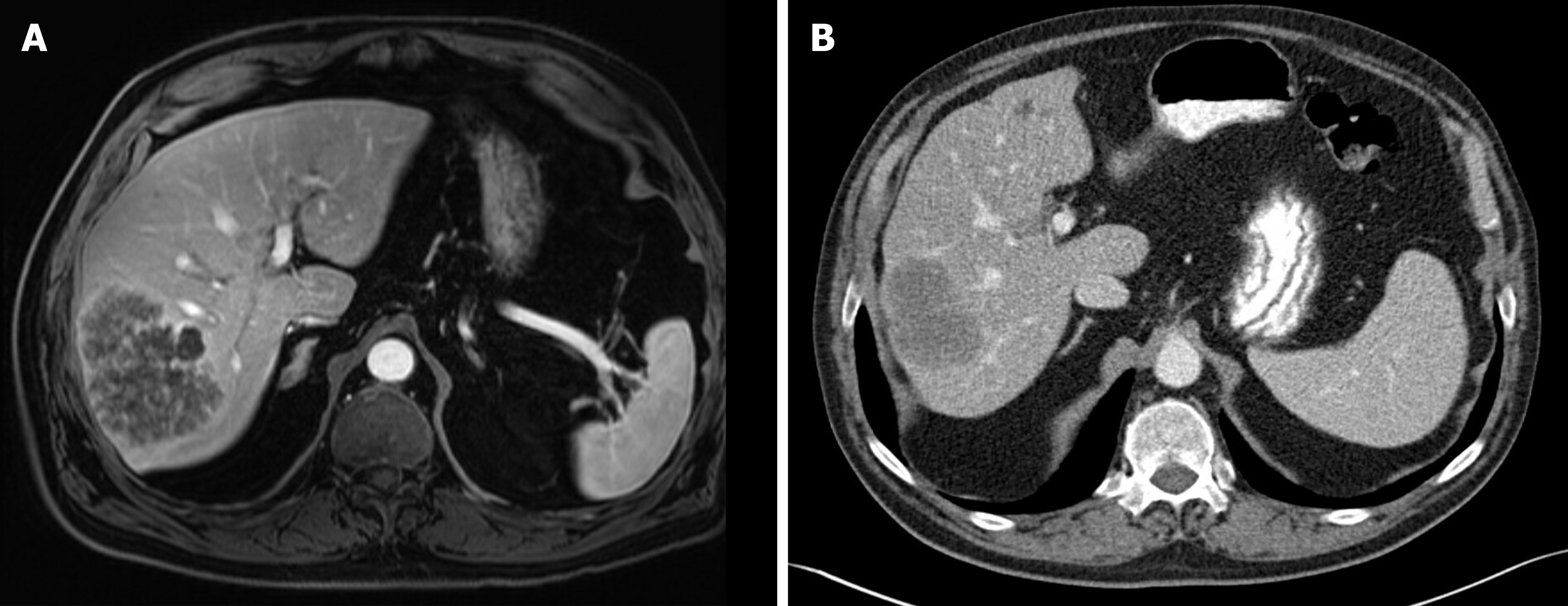
Figure 1 Radiographic response to neoadjuvant chemotherapy.
A: A 70-year-old man presented with a T2-hyperintense metachronous rectal liver metastasis, seen on magnetic resonance imaging; B: Computed tomography scan performed after 6 cycles of FOLFOX plus bevacizumab showed partial Response Evaluation Criteria in Solid Tumors response with decrease in maximum diameter from 9.6 cm to 7.0 cm, as well as optimal morphologic response. The section planes displayed above show the tumor in maximum diameter. The patient subsequently underwent right posterior sectionectomy with no evidence of disease 3 yr after surgery.
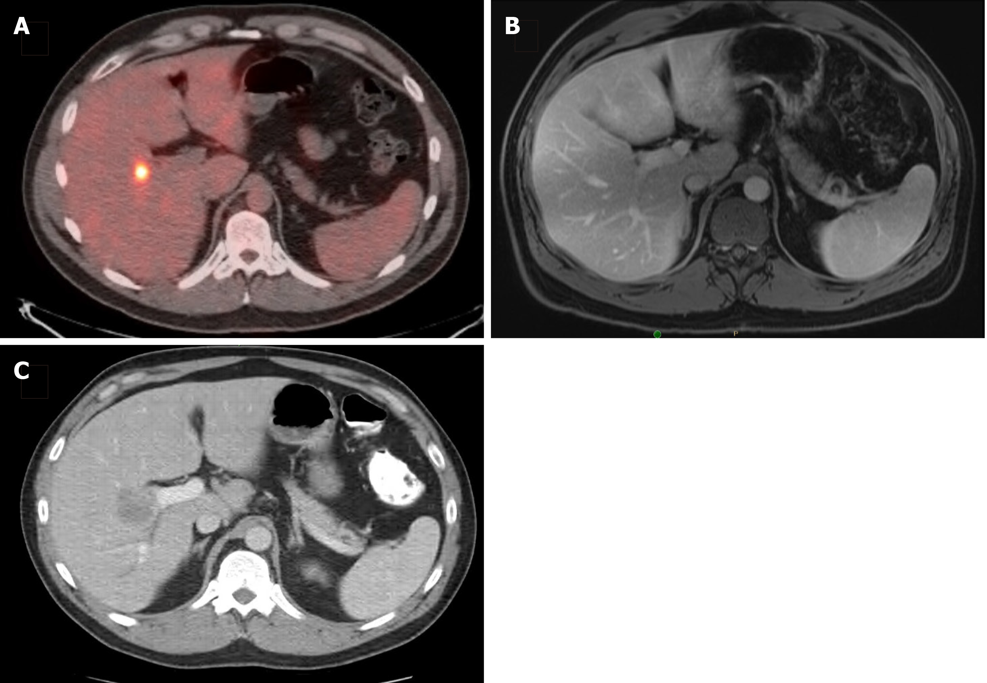
Figure 2 Disappearing liver metastasis.
A: A 36-year-old man presented with a solitary synchronous colorectal liver metastasis to segment 5, seen on staging positron emission tomography-computed tomography scan; B: After 6 cycles of CAPOX plus bevacizumab, followed by pelvic radiation (5040 cGy in 28 fractions), subsequent magnetic resonance imaging (shown) and intraoperative ultrasound were unable to localize the lesion; C: He was then lost to follow-up and returned 18 mo later with an intrahepatic recurrence, seen on surveillance computed tomography scan and confirmed with tissue biopsy.
- Citation: Guo M, Jin N, Pawlik T, Cloyd JM. Neoadjuvant chemotherapy for colorectal liver metastases: A contemporary review of the literature. World J Gastrointest Oncol 2021; 13(9): 1043-1061
- URL: https://www.wjgnet.com/1948-5204/full/v13/i9/1043.htm
- DOI: https://dx.doi.org/10.4251/wjgo.v13.i9.1043


