Copyright
©The Author(s) 2021.
World J Gastrointest Oncol. Mar 15, 2021; 13(3): 174-184
Published online Mar 15, 2021. doi: 10.4251/wjgo.v13.i3.174
Published online Mar 15, 2021. doi: 10.4251/wjgo.v13.i3.174
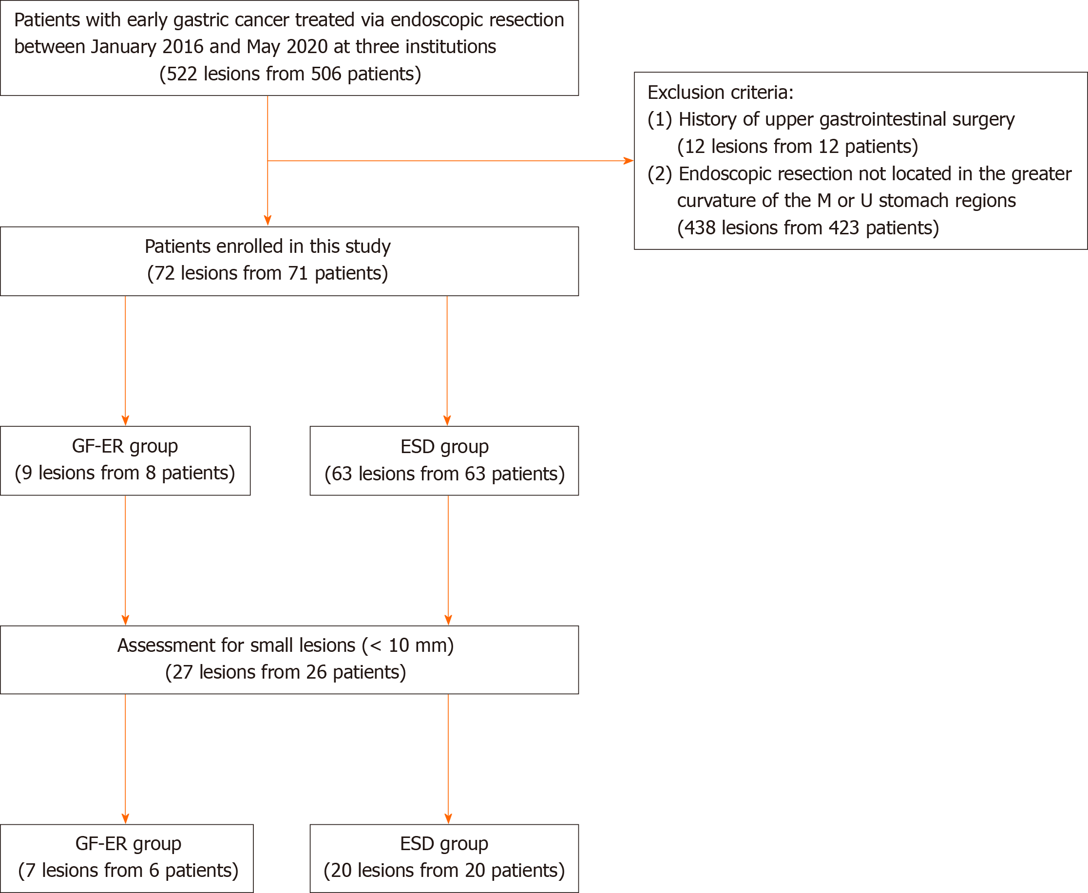
Figure 1 Study flowchart.
ESD: Endoscopic submucosal dissection; GF-ER: Grasping forceps-assisted endoscopic resection; M: Middle; U: Upper.
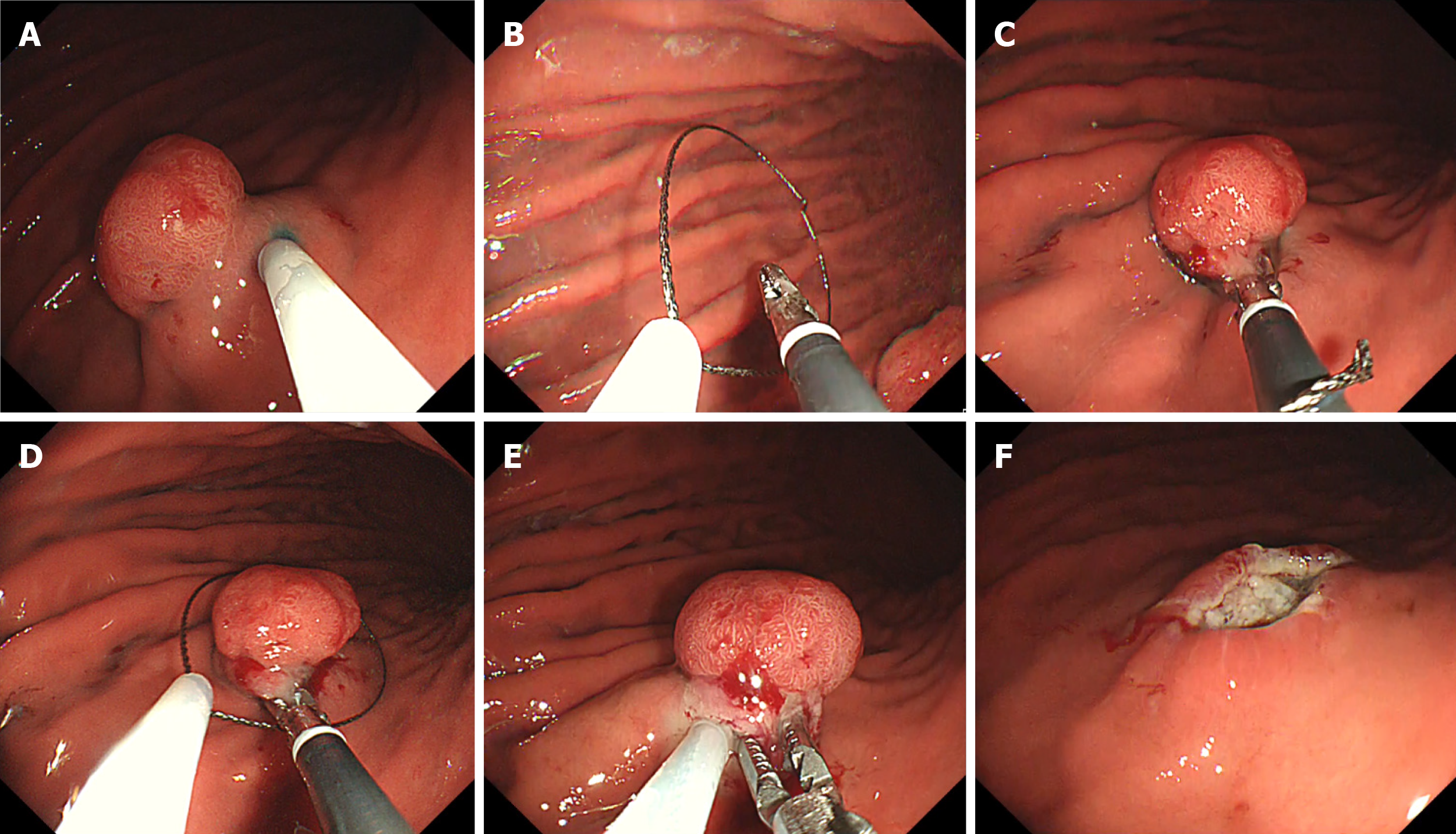
Figure 2 The grasping forceps-assisted endoscopic resection procedure.
A: Normal saline solution was injected into the submucosa around the lesion; B: A snare and grasping snare were both deployed through one of the scope’s two channels; C: The grasping snare was used to firmly grasp the elevated mucosa; D: The snare encircled the grasped mucosa; E: We ensured that the entire lesion was inside the snare, and then the resection was performed; F: After the resection, the mucosal defect was checked for residual tumour.
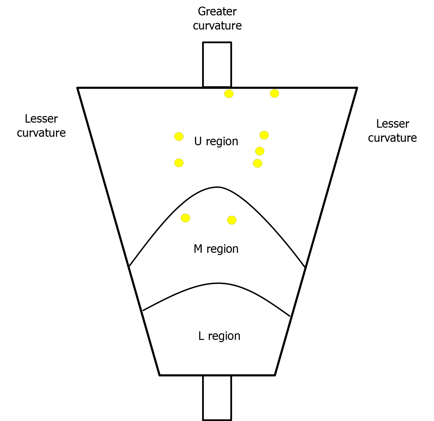
Figure 3 Location mapping for the 9 cases of early gastric cancer treated using grasping forceps-assisted endoscopic resection.
The yellow circles show the locations of the lesions that were treated using grasping forceps-assisted endoscopic resection. L: Lower; M: Middle; U: Upper.
- Citation: Ichijima R, Suzuki S, Esaki M, Horii T, Kusano C, Ikehara H, Gotoda T. Efficacy and safety of grasping forceps-assisted endoscopic resection for gastric neoplasms: A multi-centre retrospective study. World J Gastrointest Oncol 2021; 13(3): 174-184
- URL: https://www.wjgnet.com/1948-5204/full/v13/i3/174.htm
- DOI: https://dx.doi.org/10.4251/wjgo.v13.i3.174


