Copyright
©The Author(s) 2021.
World J Gastrointest Oncol. Nov 15, 2021; 13(11): 1680-1695
Published online Nov 15, 2021. doi: 10.4251/wjgo.v13.i11.1680
Published online Nov 15, 2021. doi: 10.4251/wjgo.v13.i11.1680
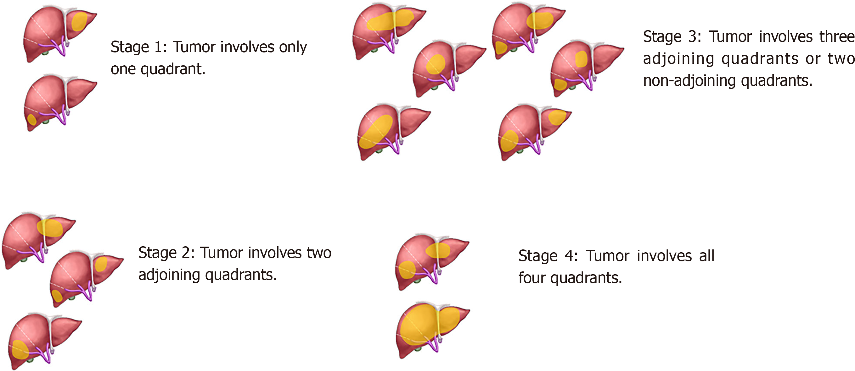
Figure 1 Definition of the pretreatment extent of disease or PRETEXT classification for the malignant liver tumor.
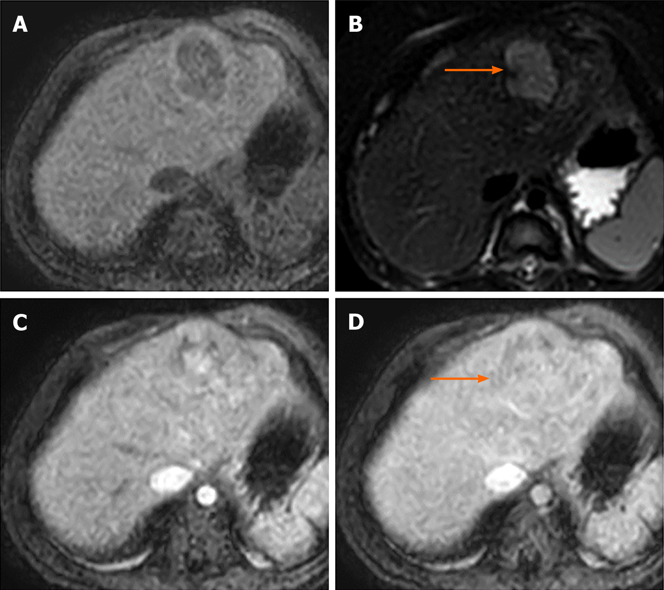
Figure 2 Magnetic resonance images of a 2-year-old boy with underlying abernethy malformation presenting with an incidentally identified liver mass with pathological tissue diagnosed hepatoblastoma.
A: Mass showing well-defined hypointense liver parenchyma on T1W and; B: Hyperintense parenchyma on T2W images; C: This mass revealed heterogeneous arterial hyperenhancement; and D: Venous enhancement after the administration of gadolinium-based contrast agent.
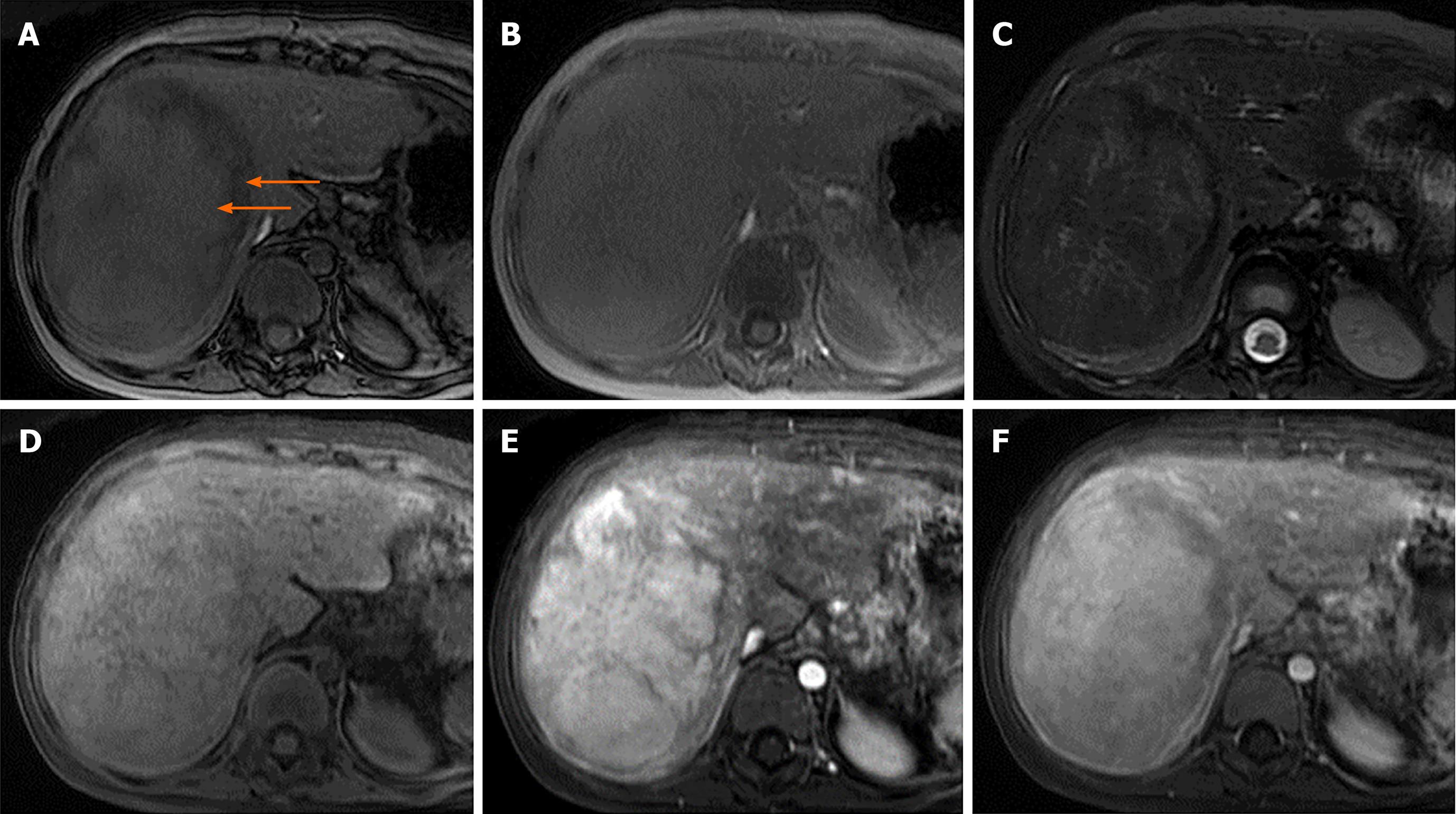
Figure 3 Magnetic resonance images of a 10-year-old girl with extrahepatic hypertension from portal vein thrombosis status post-splenectomy and proximal splenorenal shunt with developing liver mass with pathological tissue diagnosis of hepatic adenoma.
A: Axial dual gradient echo opposed-phase images revealed a heterogeneous drop in signal intensity (arrows); B: Axial dual gradient echo in the in-phase image revealing the heterogeneous microscopic fat in the mass; C: Heterogeneously mild hyperintense mass in T2-weighted (T2W) image; D: Iso-to-slightly hyperintense mass in the T1W image; E: Intense arterial hyperenhancement after gadolinium-based contrast administration; F: Heterogeneous venous enhancement after gadolinium-based contrast administration.
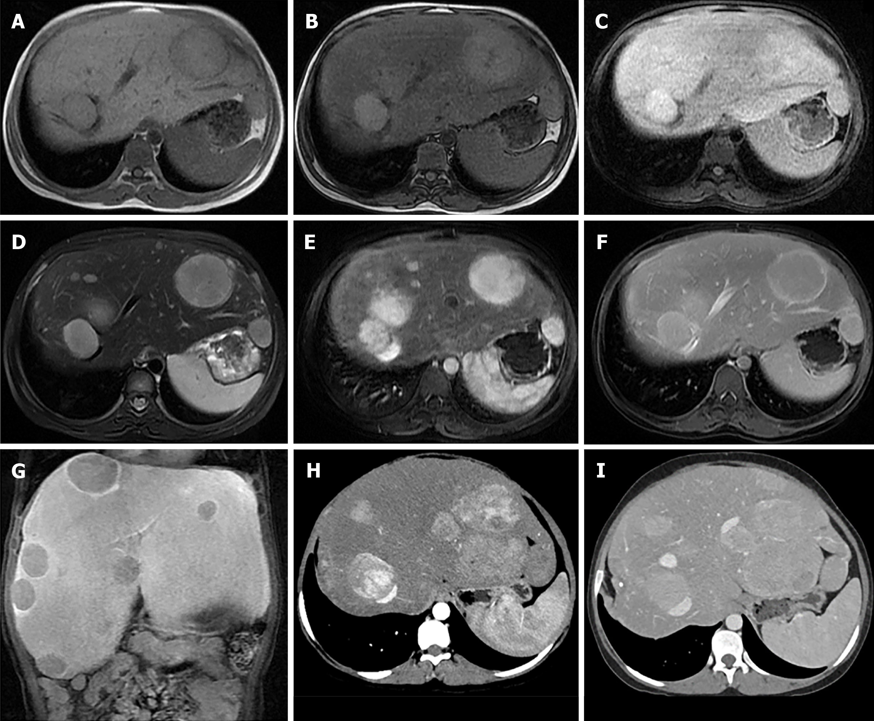
Figure 4 Magnetic resonance images of a 15-year-old girl with underlying GSD type 1 (A-G) and at the 5-yr follow-up, computed tomography was performed (H-I).
One of the identified nodules presented histopathological findings compatible with hepatic adenoma. A and B: Axial dual gradient echo images showing several slightly hyperintense nodules in both hepatic lobes on the background of diffuse hepatic steatosis with dropout in signal intensity of liver parenchyma on contrast-phase images (A) as compared to in-phase images (B); C: Hypointense nodules on the T1-weighted (T1W) image; D: Hyperintense nodules on the T2W image; E-G: Intense arterial hyperenhancement of nodules (E), iso-to mild venous enhancement (F) and hypointense nodules on delayed hepatobiliary phases at 20 min (G) after injection with gadolinium-base hepatocyte specific contrast agent; H: Intense arterial hyperenhancement and increased in size of the nodules; I: Slight hyperenhancement in the venous phase.
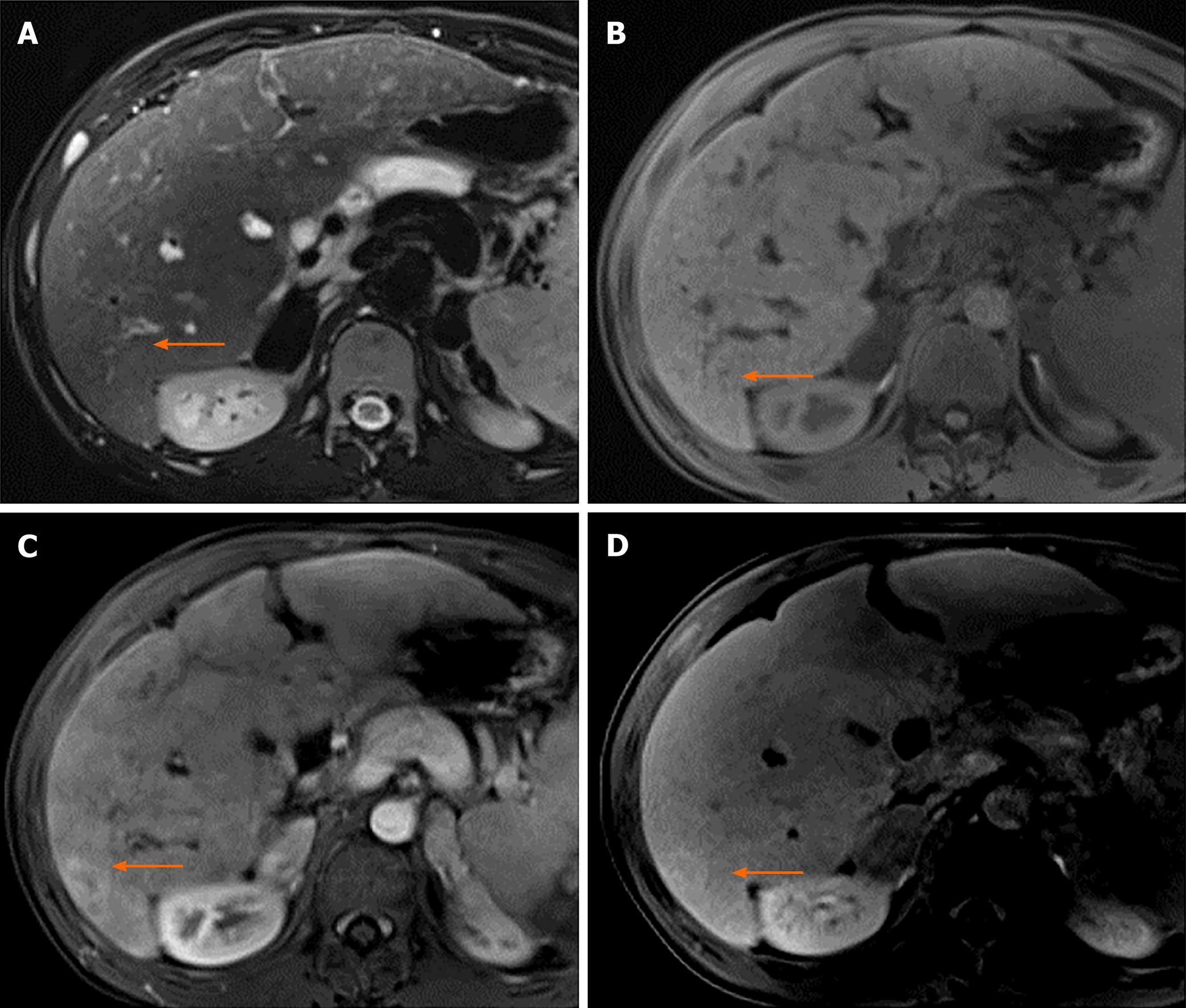
Figure 5 Pre-operative magnetic resonance images of a 13-year-old boy with biliary atresia identified liver nodule (arrows) before liver transplantation.
The histopathological findings of the nodule were compatible with FNH. A: Isointense nodule on the T2W image; B: Iso-to-slightly hyperintense nodule on the T1W image; C: Arterial hyperenhancement of the nodule and; D: Persisted delayed enhancement on delayed hepatobiliary phase at 30 min after the administration of gadolinium-based hepatocyte specific contrast agent.
Figure 6 Histopathology from liver tumor demonstrates hepatocellular carcinoma.
A: Hematoxylin and eosin staining shows tumor cells in a trabecular pattern. The cell plates are three cells thick or wider in most of this tumor; B: Reticulin staining highlights loss of normal cord architecture; C: Cluster of differentiation 34 immunostaining highlights the increased vascularity of hepatocellular carcinoma.
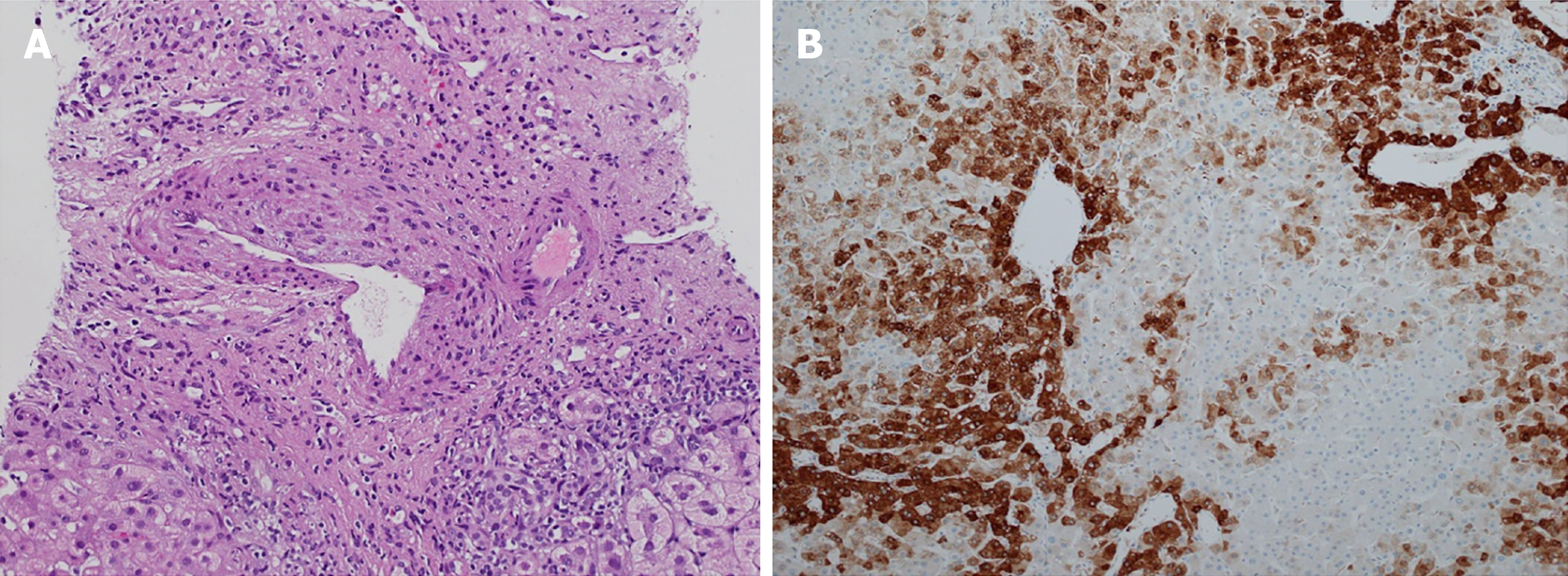
Figure 7 Histopathology of the liver tumor demonstrates focal nodular hyperplasia.
A: Hematoxylin and eosin staining reveals a muscular vessel with irregular wall thickness, and ductular reaction; B: Glutamine synthetase immunostaining of focal nodular hyperplasia shows increased overall staining in a map-like pattern.
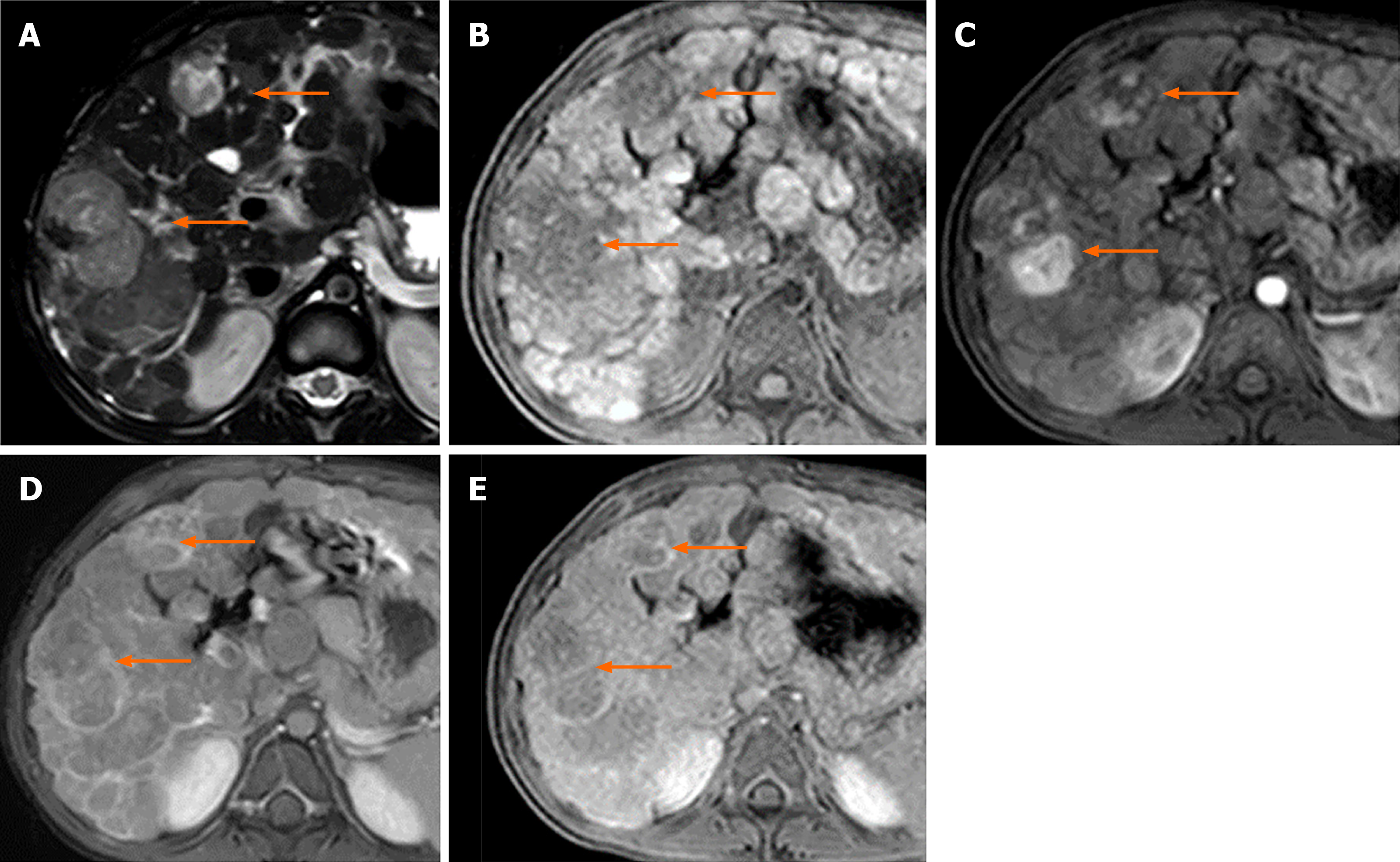
Figure 8 Magnetic resonance images of a 7-yr-old boy with tyrosinemia type I and renal Fanconi syndrome presenting nodules with histopathologic findings indicative of hepatocellular carcinoma.
He underwent liver transplantation with favorable outcome and no evidence of tumor recurrence after a 1-yr follow-up. A: Multiple hyperintense nodules in T2-weighted (T2W) image; B: Hypointense nodules on the T1W image on the background of macronodular cirrhotic liver; C: Heterogeneous venous “wash-out” enhancement of the nodules; and D: Delayed capsular enhancement after administration of gadolinium-based contrast agent.
- Citation: Sintusek P, Phewplung T, Sanpavat A, Poovorawan Y. Liver tumors in children with chronic liver diseases. World J Gastrointest Oncol 2021; 13(11): 1680-1695
- URL: https://www.wjgnet.com/1948-5204/full/v13/i11/1680.htm
- DOI: https://dx.doi.org/10.4251/wjgo.v13.i11.1680


