Copyright
©The Author(s) 2020.
World J Gastrointest Oncol. Nov 15, 2020; 12(11): 1372-1380
Published online Nov 15, 2020. doi: 10.4251/wjgo.v12.i11.1372
Published online Nov 15, 2020. doi: 10.4251/wjgo.v12.i11.1372
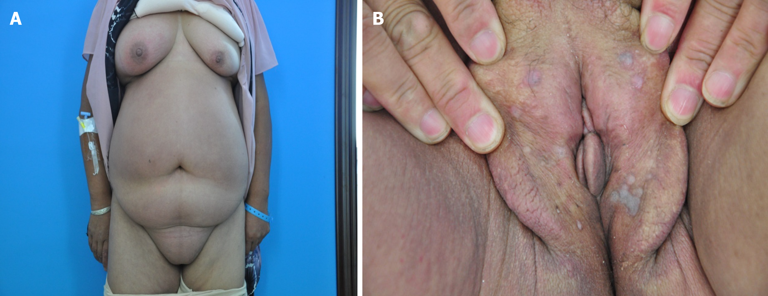
Figure 1 Photographs of our patient.
A: Diffuse erythema and swelling on the chest, abdomen, and right leg; B: Edema and a number of flat skin-colored or gray papules on the labia majora.
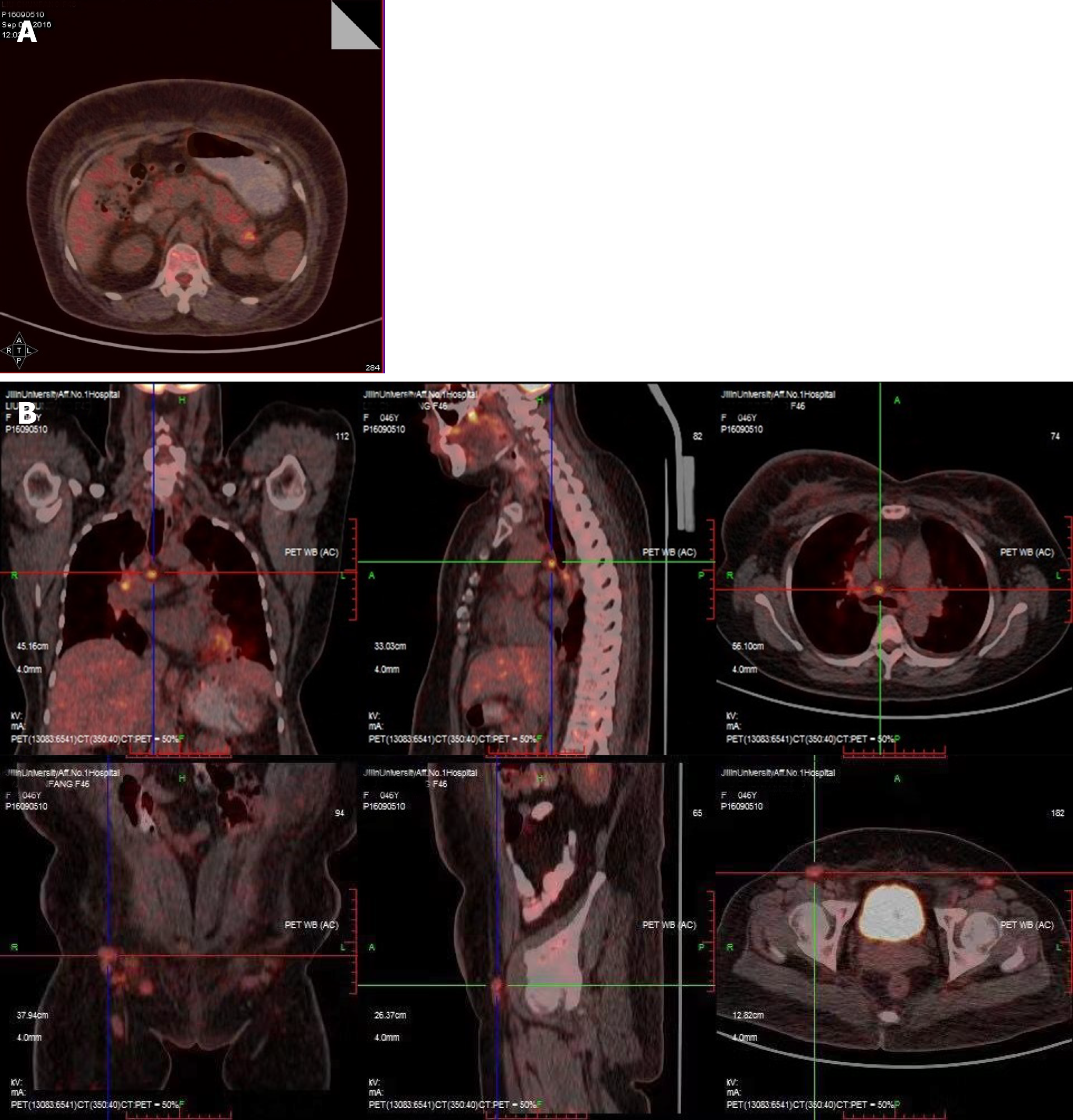
Figure 2 Positron emission tomography-computed tomography scan showing increased metabolic activity.
A: The tail of the pancreas; B: Mediastinum, hilus of the lung, and postperitoneal lymph nodes.
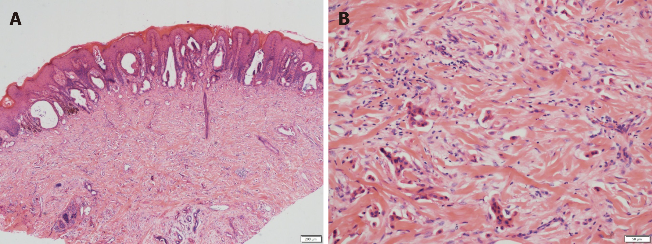
Figure 3 Pathology of cutaneous metastasis.
A: Histological examination of the labia majora papule showing nests of moderately differentiated atypical cells partly forming adenomatous structures in the collagen bundle of the dermis and lymphangiectasis in the dermis [hematoxylin-eosin (HE) staining, × 40]; B: Dermis occupied by mass of numerous tumor cells (HE staining, × 200).
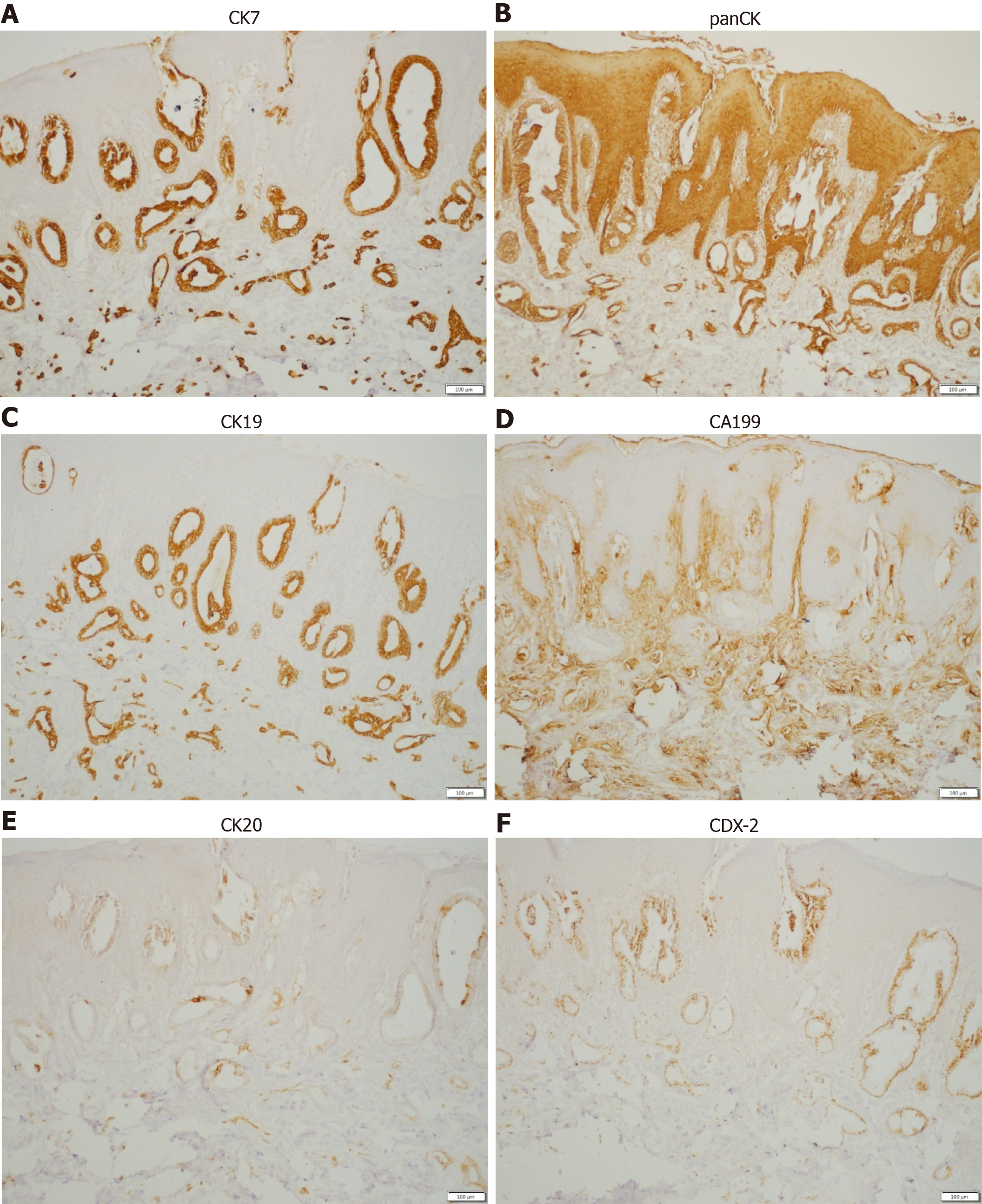
Figure 4 Neoplastic glands showing a positive reaction to immunohistochemical staining (100 ×).
A: CK7 (+); B: panCK (+); C: CK19 (+); D: CA199 (+); E: CK20 (+); F: CDX-2 (+).
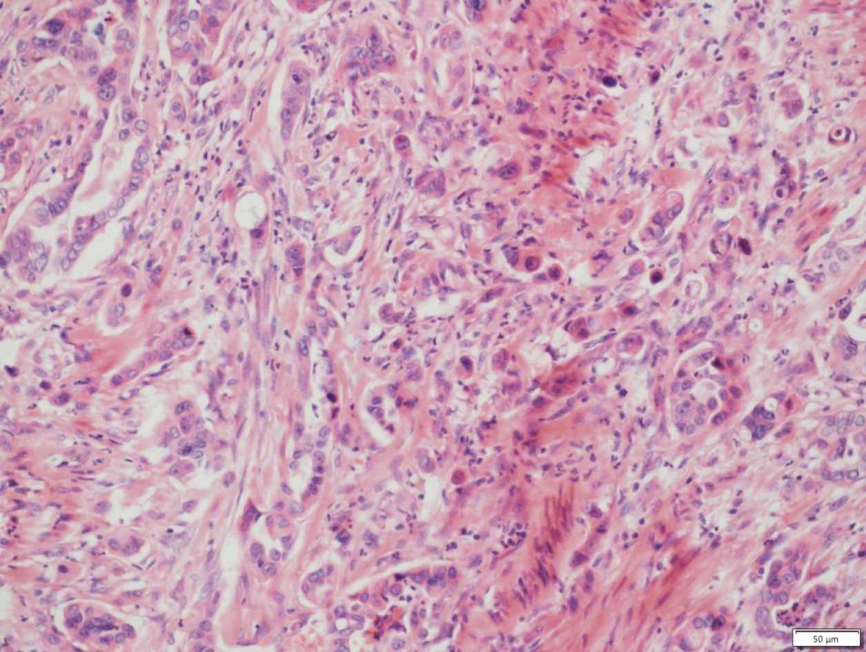
Figure 5 B-mode ultrasound-guided needle biopsy of the pancreas showing the moderately and poorly differentiated adenocarcinoma (hematoxylin-eosin staining, × 200).
- Citation: Shi Y, Li SS, Liu DY, Yu Y. Cutaneous metastases of pancreatic carcinoma to the labia majora: A case report and review of literature. World J Gastrointest Oncol 2020; 12(11): 1372-1380
- URL: https://www.wjgnet.com/1948-5204/full/v12/i11/1372.htm
- DOI: https://dx.doi.org/10.4251/wjgo.v12.i11.1372


