Copyright
©The Author(s) 2020.
World J Gastrointest Oncol. Nov 15, 2020; 12(11): 1364-1371
Published online Nov 15, 2020. doi: 10.4251/wjgo.v12.i11.1364
Published online Nov 15, 2020. doi: 10.4251/wjgo.v12.i11.1364
Figure 1 Colonoscopy showed abnormal colonic edematous mucosa with multiple ulcers from the sigmoid colon, distal to the anastomotic site, to the rectosigmoid colon and rectum below the peritoneal reflection.
A: Mild inflammation in the anastomotic site; B: Severe inflammation with multiple ulcers and edematous mucosa in the sigmoid colon; C: Rectum below the peritoneal reflection with almost normal findings.
Figure 2 Abdominal contrast-enhanced computed tomography at 5 year after left hemicolectomy and at 1 year after the start of palliative chemotherapy.
A: Computed tomography (CT) showed edematous change from the sigmoid colon to the rectum (triple arrow); B: An aneurysm adjacent to the inferior mesenteric vein (IMV) (arrow); C: Multidetector CT angiography (three-dimensional volume-rendered image) showed the arteriovenous fistula with nidus involving the branches of the inferior mesenteric artery and IMV.
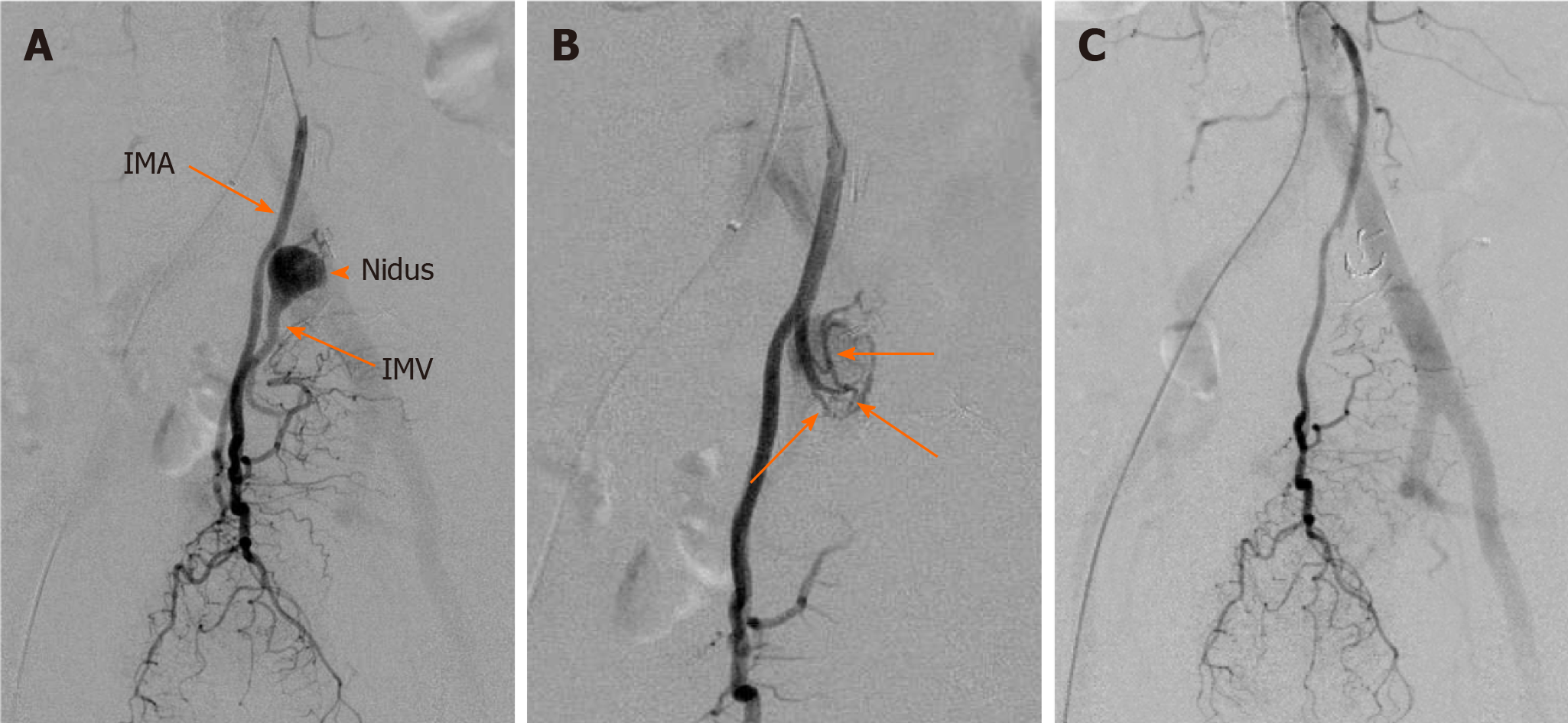
Figure 3 An elective angiography of the inferior mesenteric artery.
A: Angiography revealed the nidus of fistula supplied by the inferior mesenteric artery (IMA) (arrow head); B: Several shunting points of the fistula from the branch of the IMA (arrows), which confirmed the existence of an inferior mesenteric arteriovenous fistula (IMAVF); C: The IMAVF completely disappeared after arterial embolization. IMV: Inferior mesenteric vein.
- Citation: Doi A, Takeda H, Umemoto K, Oumi R, Wada S, Hamaguchi S, Mimura H, Arai H, Horie Y, Mizukami T, Izawa N, Ogura T, Nakajima TE, Sunakawa Y. Inferior mesenteric arteriovenous fistula during treatment with bevacizumab in colorectal cancer patient: A case report. World J Gastrointest Oncol 2020; 12(11): 1364-1371
- URL: https://www.wjgnet.com/1948-5204/full/v12/i11/1364.htm
- DOI: https://dx.doi.org/10.4251/wjgo.v12.i11.1364


