Copyright
©The Author(s) 2018.
World J Gastrointest Oncol. Dec 15, 2018; 10(12): 516-521
Published online Dec 15, 2018. doi: 10.4251/wjgo.v10.i12.516
Published online Dec 15, 2018. doi: 10.4251/wjgo.v10.i12.516
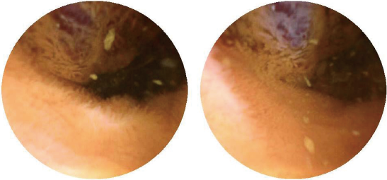
Figure 1 Capsule endoscopic appearance of the lesion.
Capsule endoscopy showed a prominent polypoid lesion in the ileum.
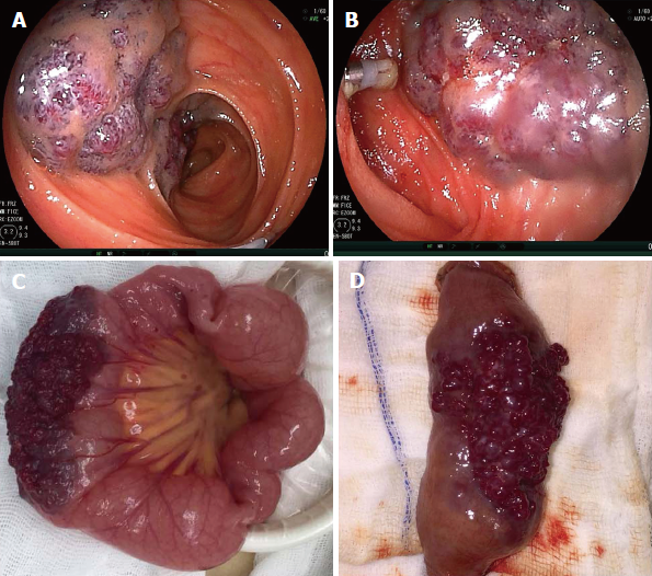
Figure 2 Endoscopic and gross appearance of the lesion.
A: Transanal double-balloon enteroscopy revealed a reddish purple lesion in the ileum about 80 cm proximal to the ileocecal valve, and a titanium clip was used to mark the limit reached; B: Transoral double-balloon enteroscopy showed the same lesion and the marked titanium clip; C: Gross intraoperative appearance of the lesion; D: Gross appearance of the lesion after resection.
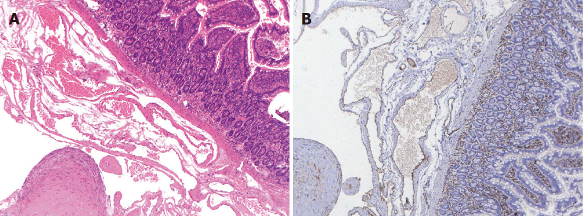
Figure 3 Histopathological examination of the lesion.
A: Hematoxylin-eosin staining showed a blood-filled sinus-like space in the whole layer of the ileum (× 50); B: Immunohistochemistry indicated the cells lined with the vascular spaces were CD31-positive (× 50).
- Citation: Hu PF, Chen H, Wang XH, Wang WJ, Su N, Shi B. Small intestinal hemangioma: Endoscopic or surgical intervention? A case report and review of literature. World J Gastrointest Oncol 2018; 10(12): 516-521
- URL: https://www.wjgnet.com/1948-5204/full/v10/i12/516.htm
- DOI: https://dx.doi.org/10.4251/wjgo.v10.i12.516


