Copyright
©The Author(s) 2018.
World J Gastrointest Oncol. Nov 15, 2018; 10(11): 370-380
Published online Nov 15, 2018. doi: 10.4251/wjgo.v10.i11.370
Published online Nov 15, 2018. doi: 10.4251/wjgo.v10.i11.370
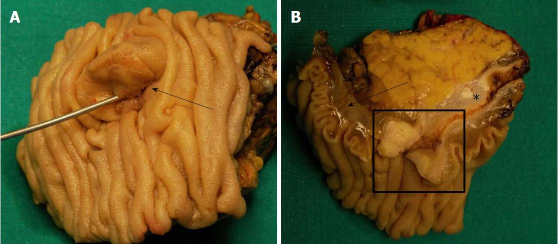
Figure 1 A classic example of the macroscopic appearance of a case of ampulla of Vater carcinoma.
A: The ampullary area is markedly enlarged (black arrow); B: On the section surface, the ampulla of Vater carcinoma (black box), the adjacent duodenal wall (black arrow) and bile duct (asterisk) are clearly visible.
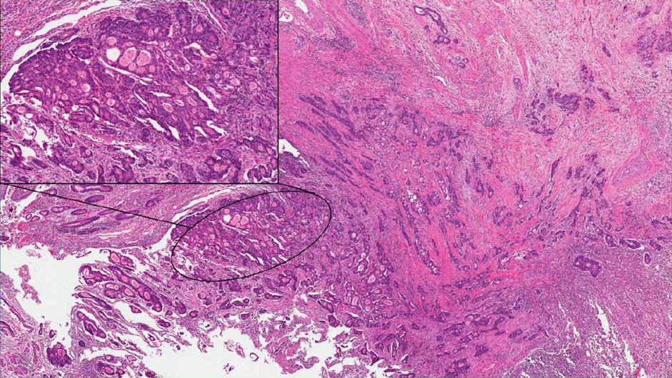
Figure 2 A classic example of intestinal-type ampulla of Vater carcinoma.
At low magnification (2 × original magnification) and at higher magnification (the box in the upper left corner, 10 × original magnification) to better show its histological features. The lesion is composed of a colorectal-like architecture, with glands characterized by comedo-like necrosis.
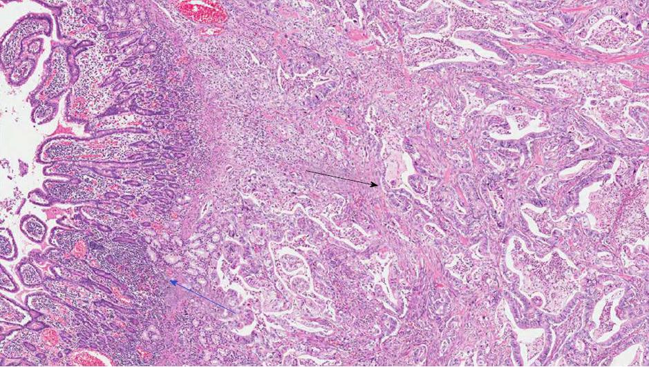
Figure 3 A classic example of pancreaticobiliary-type ampulla of Vater carcinoma (original magnification: 20 ×).
The lesion is composed of ductal adenocarcinoma-like glands (black arrow) invading the duodenum (blue arrow).
Figure 4 Immunohistochemical analysis of an ampullary adenocarcinoma of mixed subtype (original magnification 20 ×).
A: Immunohistochemical analysis of an ampullary adenocarcinoma of mixed subtype, with cytokeratin 20 (CK20); B: Immunohistochemical analysis of an ampullary adenocarcinoma of mixed subtype, with cytokeratin 7 (CK7). This image highlights that, in the same area, some neoplastic glands may be positive not only for CK7 or for CK20, but for both markers even. The coexpression of an intestinal marker, such as CK20, and of a pancreatobiliary marker, such as CK7, supports the classification as mixed subtype.
- Citation: Pea A, Riva G, Bernasconi R, Sereni E, Lawlor RT, Scarpa A, Luchini C. Ampulla of Vater carcinoma: Molecular landscape and clinical implications. World J Gastrointest Oncol 2018; 10(11): 370-380
- URL: https://www.wjgnet.com/1948-5204/full/v10/i11/370.htm
- DOI: https://dx.doi.org/10.4251/wjgo.v10.i11.370


