Copyright
©2013 Baishideng Publishing Group Co.
World J Gastrointest Endosc. Jul 16, 2013; 5(7): 313-322
Published online Jul 16, 2013. doi: 10.4253/wjge.v5.i7.313
Published online Jul 16, 2013. doi: 10.4253/wjge.v5.i7.313
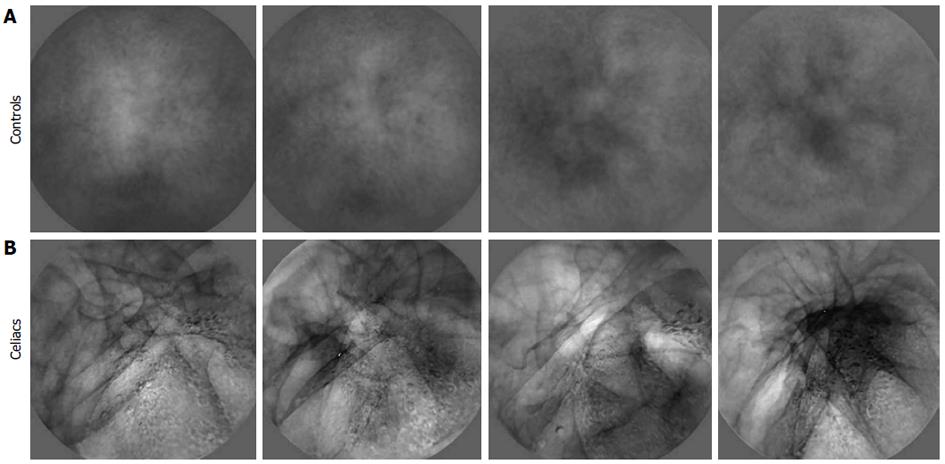
Figure 4 Examples of basis images derived from celiac vs control videoclip series.
A: Control; B: Celiacs. The celiac basis has more heterogeneous structure. There are dark lines running through the celiac basis images as well as black and white blotches. In contrast, the control basis images are mostly uniform, with only diffuse features being evident.
- Citation: Ciaccio EJ, Tennyson CA, Bhagat G, Lewis SK, Green PH. Implementation of a polling protocol for predicting celiac disease in videocapsule analysis. World J Gastrointest Endosc 2013; 5(7): 313-322
- URL: https://www.wjgnet.com/1948-5190/full/v5/i7/313.htm
- DOI: https://dx.doi.org/10.4253/wjge.v5.i7.313


