Copyright
©The Author(s) 2017.
World J Gastrointest Endosc. Jun 16, 2017; 9(6): 267-272
Published online Jun 16, 2017. doi: 10.4253/wjge.v9.i6.267
Published online Jun 16, 2017. doi: 10.4253/wjge.v9.i6.267
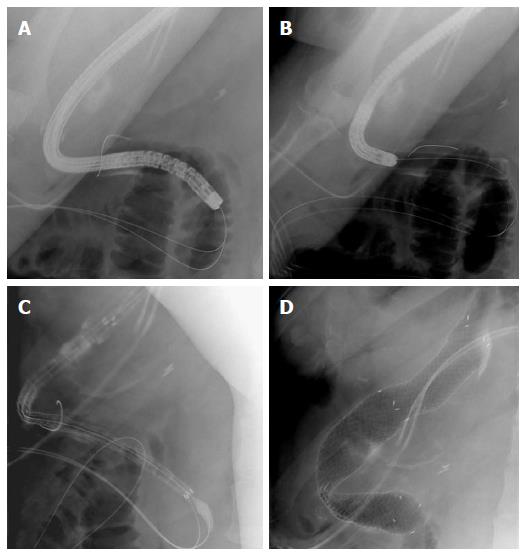
Figure 1 Deployement of the bariatric stent.
A: Fluoroscopic image showing advancement of the overtube and endoscope through the sleeve gastrectomy; B: Fluoroscopic image depicting removal of the endoscope after placement of the guidewire and the overtube; C: Fluoroscopic image revealing the progression of the stent over-the-wire and through the overtube; D: Fluoroscopic image showing the release of the stent after the overtube was slightly pulled back. A marked angulation of the stent is seen.
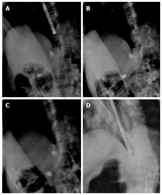
Figure 2 Deployement of the bariatric stent.
A: Fluoroscopic image showing the overtube correctly placed in the sleeve gastrectomy while the endoscope is being removed; B: Fluoroscopic image depicting the overtube and the guidewire in place; C: Fluoroscopic image revealing the advancement of the stent over-the-wire and through the overtube; D: Fluoroscopic image showing the deployed stent with a marked angulation.
- Citation: Ponte A, Pinho R, Proença L, Silva J, Rodrigues J, Sousa M, Silva JC, Carvalho J. Utility of the balloon-overtube-assisted modified over-the-wire stenting technique to treat post-sleeve gastrectomy complications. World J Gastrointest Endosc 2017; 9(6): 267-272
- URL: https://www.wjgnet.com/1948-5190/full/v9/i6/267.htm
- DOI: https://dx.doi.org/10.4253/wjge.v9.i6.267


