Copyright
©The Author(s) 2017.
World J Gastrointest Endosc. Feb 16, 2017; 9(2): 70-76
Published online Feb 16, 2017. doi: 10.4253/wjge.v9.i2.70
Published online Feb 16, 2017. doi: 10.4253/wjge.v9.i2.70
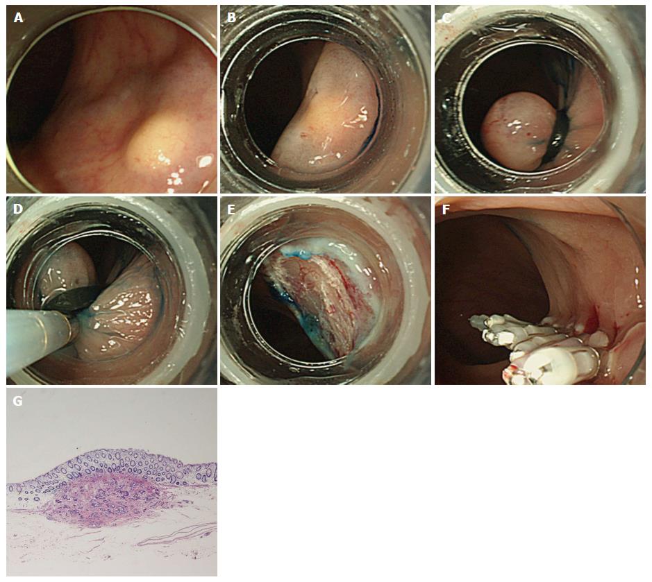
Figure 1 Endoscopic submucosal resection with a ligation device.
A: Endoscopic view of a carcinoid tumor in the rectum; B: Submucosal injection beneath the tumor with glycerin solution; C: An elastic band was deployed, and then pseudopolyp was created; D: Snare resection was performed beneath the elastic band; E: An artificial ulcer was observed; F: Endoscopic plication was performed with the use of metal endoclips; G: Histopathological examination showed en bloc resection of the carcinoid tumor.
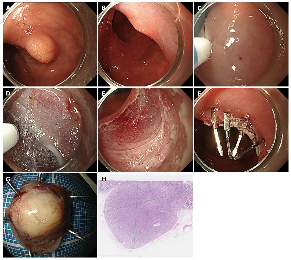
Figure 2 Endoscopic submucosal dissection.
A: Endoscopic view of a carcinoid tumor in the rectum; B: Submucosal injection beneath the tumor with sodium hyaluronate; C: A hemicircumferential incision was performed with the use of the electrosurgical knife; D: A submucosal pocket was created during ESD. Submucosal dissection was performed just above the muscular layer; E: An artificial ulcer was observed; F: Endoscopic plication was performed with the use of metal endoclips; G: The specimen resected by ESD; H: Histopathological examination showed en bloc resection of the carcinoid tumor. ESD: Endoscopic submucosal dissection.
- Citation: Harada H, Suehiro S, Murakami D, Nakahara R, Shimizu T, Katsuyama Y, Miyama Y, Hayasaka K, Tounou S. Endoscopic submucosal dissection for small submucosal tumors of the rectum compared with endoscopic submucosal resection with a ligation device. World J Gastrointest Endosc 2017; 9(2): 70-76
- URL: https://www.wjgnet.com/1948-5190/full/v9/i2/70.htm
- DOI: https://dx.doi.org/10.4253/wjge.v9.i2.70


