Copyright
©The Author(s) 2017.
World J Gastrointest Endosc. Dec 16, 2017; 9(12): 579-582
Published online Dec 16, 2017. doi: 10.4253/wjge.v9.i12.579
Published online Dec 16, 2017. doi: 10.4253/wjge.v9.i12.579
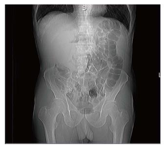
Figure 1 Abdominal radiography showing right upper quadrant mass enlargement.
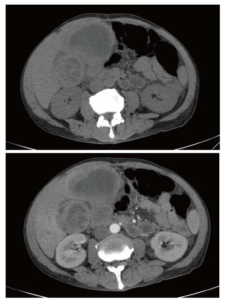
Figure 2 Computed tomography scan (simple and arterial phase) with infiltrative lesion at duodenum and gallbladder.
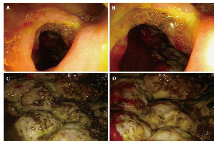
Figure 3 Duodenal posterior wall perforation (A and B), retroperitoneal solid and irregular neoplasia (C), extreme friability and spontaneous bleeding (D).
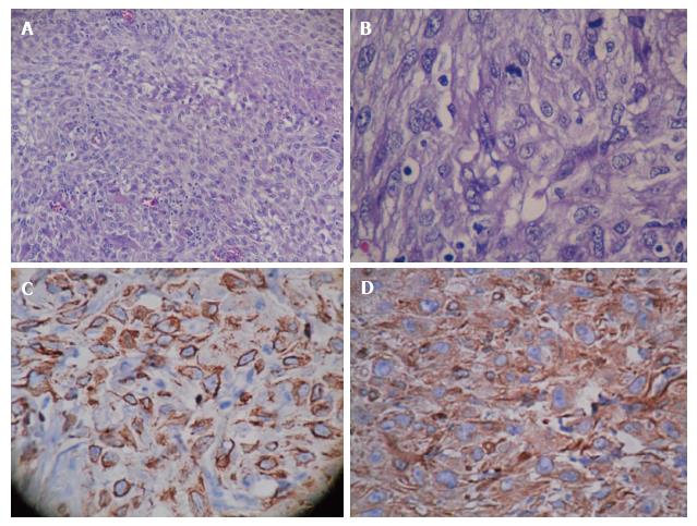
Figure 4 H and E with fusiform and giant pleomorphic cells, increased mitosis (A and B), positive staining for vimentin and cytokeratin respectively (C and D).
- Citation: Coronado JA, Chávez MÁ, Manrique MA, Cerna J, Trejo AL. Retroperitoneal epithelioid sarcoma: A case report. World J Gastrointest Endosc 2017; 9(12): 579-582
- URL: https://www.wjgnet.com/1948-5190/full/v9/i12/579.htm
- DOI: https://dx.doi.org/10.4253/wjge.v9.i12.579


