Copyright
©The Author(s) 2016.
World J Gastrointest Endosc. Oct 16, 2016; 8(18): 653-662
Published online Oct 16, 2016. doi: 10.4253/wjge.v8.i18.653
Published online Oct 16, 2016. doi: 10.4253/wjge.v8.i18.653
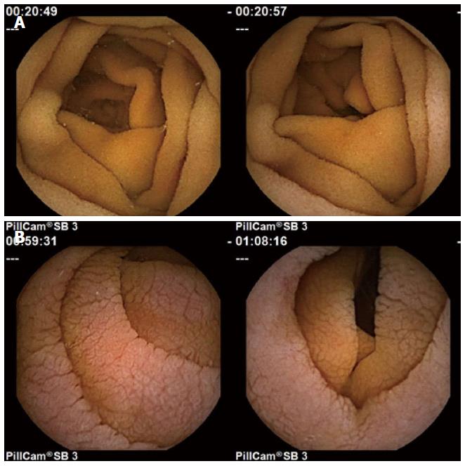
Figure 1 Normal (A) vs untreated celiac patient images (B).
Note the presence of mucosal folds, a mottled appearance, and fissuring, in the images from untreated celiac patients (lower).
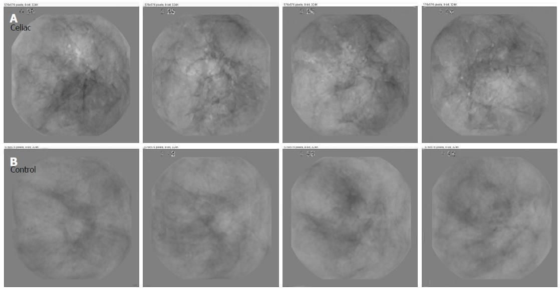
Figure 2 Evidence of more prominent fissuring is present in untreated control images (B) as compared with celiac basis images (A).
The basis images are modified from a series of original endoscopic images so that salient features are enhanced.
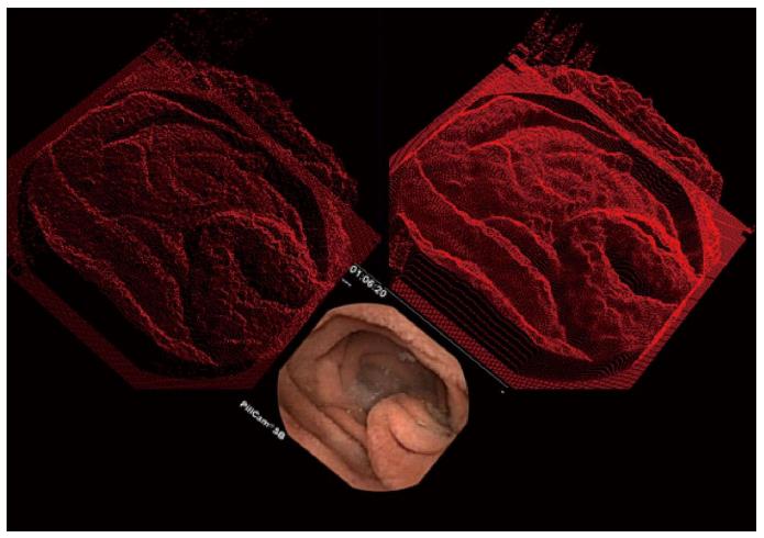
Figure 3 Shape-from-shading is used to render two-dimensional endoscopic images (lower panel) into three-dimensional constructs.
First the color image is converted to grayscale. Then the degree of pixel brightness is linearly interpreted as depth in the constructs at top. Lighter areas in the lower image appear as taller protrusions in the images at top. Top left: Half resolution three-dimensional reconstruction; Top right: Full resolution three-dimensional reconstruction.
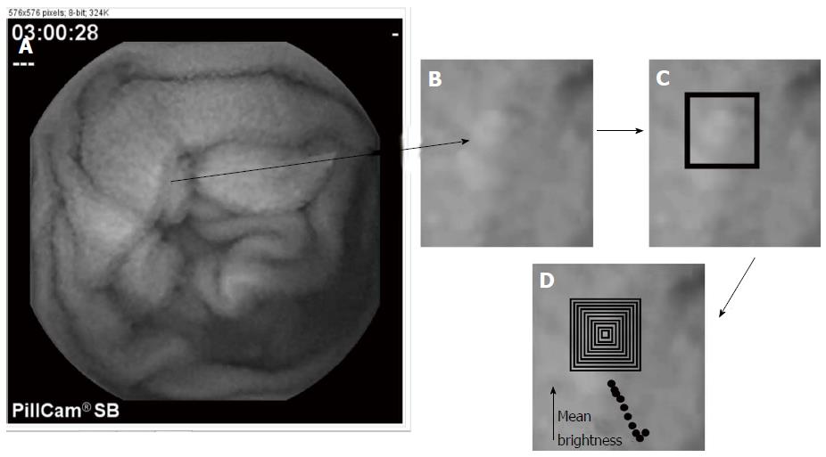
Figure 4 Example of the construction of a syntactic prototypical template.
A: Video capsule endoscopic image in grayscale; B: Area with bright center is noted by the arrow; C: A square is used to identify the area of a protrusion; D: The series of concentric squares (rings) are used to determine the protrusion dimensions. The average pixel brightness diminishes from center square to outer square. The width and length of the protrusion is defined based on the outermost square, which is that square after which a larger concentric outer square would have an increased brightness. The height of the protrusion is the difference in brightness level from its center to its outermost concentric square.
- Citation: Ciaccio EJ, Bhagat G, Lewis SK, Green PH. Recommendations to quantify villous atrophy in video capsule endoscopy images of celiac disease patients. World J Gastrointest Endosc 2016; 8(18): 653-662
- URL: https://www.wjgnet.com/1948-5190/full/v8/i18/653.htm
- DOI: https://dx.doi.org/10.4253/wjge.v8.i18.653


