Copyright
©The Author(s) 2016.
World J Gastrointest Endosc. Jul 25, 2016; 8(14): 496-500
Published online Jul 25, 2016. doi: 10.4253/wjge.v8.i14.496
Published online Jul 25, 2016. doi: 10.4253/wjge.v8.i14.496
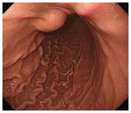
Figure 1 Gastrodudenal endoscopy showing a 5-cm firm non pulsating sub mucosal lesion in the fundus.
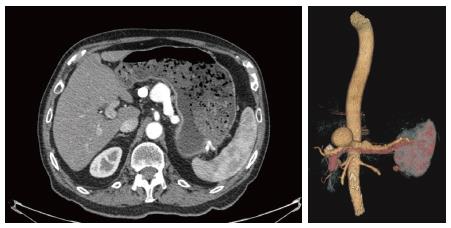
Figure 2 Abdominal enhanced computed tomography scan and angioscan showing a double aneurysm of splenic artery.
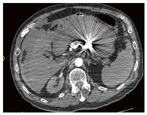
Figure 3 Abdominal enhanced computed tomography scan showing a peritoneal leak of contrast material from splenic aneurysm.
- Citation: Tannoury J, Honein K, Abboud B. Splenic artery aneurysm presenting as a submucosal gastric lesion: A case report. World J Gastrointest Endosc 2016; 8(14): 496-500
- URL: https://www.wjgnet.com/1948-5190/full/v8/i14/496.htm
- DOI: https://dx.doi.org/10.4253/wjge.v8.i14.496


