Copyright
©The Author(s) 2016.
World J Gastrointest Endosc. Jul 10, 2016; 8(13): 472-476
Published online Jul 10, 2016. doi: 10.4253/wjge.v8.i13.472
Published online Jul 10, 2016. doi: 10.4253/wjge.v8.i13.472
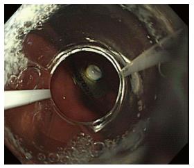
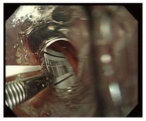
Figure 1 Endoscopic image of the push-through pack in the stomach.
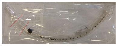
Figure 2 An endotracheal tube was cut into three sections, and the middle section (white arrow) was used as an endoscopic hood.
The red lines show the locations of the cuts made in the tube.
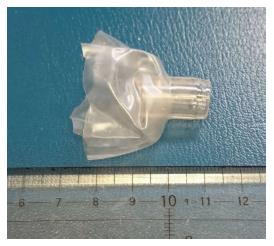
Figure 3 An endoscopic hood made from the cut endotracheal tube hood.
The middle section of the cut endotracheal tube is shown in Figure 1.
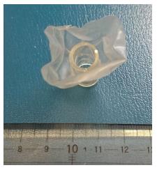
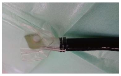
Figure 4 The cut cuff used to cloak the push-through pack was approximately 30 mm in diameter.
Figure 5 An endoscopic image.
The push-through pack in the stomach was grasped with biopsy forceps and pulled into the cut cuff, not inside the tube.
Figure 6 All four edges of the push-through pack were drawn into the cut cuff.
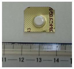
Figure 7 The size of the impacted push-through pack was 19 mm × 16 mm.
- Citation: Tateno Y, Suzuki R. Cut endotracheal tube for endoscopic removal of an ingested push-through pack. World J Gastrointest Endosc 2016; 8(13): 472-476
- URL: https://www.wjgnet.com/1948-5190/full/v8/i13/472.htm
- DOI: https://dx.doi.org/10.4253/wjge.v8.i13.472


