Copyright
©The Author(s) 2016.
World J Gastrointest Endosc. Jun 25, 2016; 8(12): 451-457
Published online Jun 25, 2016. doi: 10.4253/wjge.v8.i12.451
Published online Jun 25, 2016. doi: 10.4253/wjge.v8.i12.451
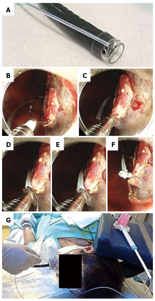
Figure 1 Clip-and-snare method using pre-looping technique.
A: The transparent cap is tightened with a snare from the outside of the endoscope (pre-looping technique) after completion of a circumferential incision; B: A tumor is seen on the lesser curvature of the upper third of the stomach. The endoscope is bent to a maximum. A hemoclip with a reusable clip deployment device is inserted through the endoscope channel and is used to grasp the edge of the tumor while avoiding its detachment from its deployment device; C, D: The pre-looped snare is loosened from the transparent cap and moved along the device toward the hemoclip; E, F: The hemoclip is tightened with the snare and released from the clip deployment device; G: The endoscopist can apply appropriate tension to the lesion using the snare independent of the endoscope. The slider of the snare is fixed with clothespins.
Figure 2 Pushing and pulling by the clip-and-snare method.
A: Clip-and-snare method using pre-looping technique (CSM-PLT) for a tumor located on the greater curvature of the middle third of the stomach; B: The endoscopist is able to obtain good visibility of the submucosal layer by pulling the snare; C: CSM-PLT was performed for another tumor located on the anterior wall of the middle third of the stomach; D: The endoscopist is able to obtain good visibility of the submucosal layer by pushing the snare (yellow arrowheads).
- Citation: Yoshida N, Doyama H, Ota R, Takeda Y, Nakanishi H, Tominaga K, Tsuji S, Takemura K. Effectiveness of clip-and-snare method using pre-looping technique for gastric endoscopic submucosal dissection. World J Gastrointest Endosc 2016; 8(12): 451-457
- URL: https://www.wjgnet.com/1948-5190/full/v8/i12/451.htm
- DOI: https://dx.doi.org/10.4253/wjge.v8.i12.451


