Copyright
©The Author(s) 2015.
World J Gastrointest Endosc. Jul 10, 2015; 7(8): 806-813
Published online Jul 10, 2015. doi: 10.4253/wjge.v7.i8.806
Published online Jul 10, 2015. doi: 10.4253/wjge.v7.i8.806
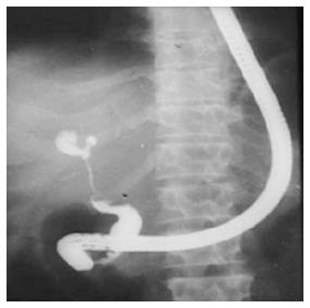
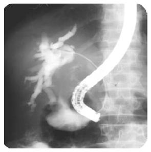
Figure 1 Postoperative type 3 hilarstricture with patent confluence.
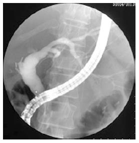
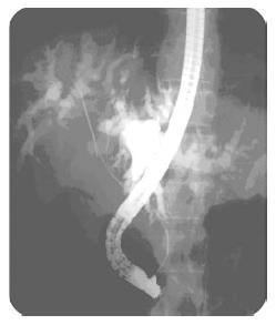
Figure 2 Postoperative type 5 hilarstricture involving right hepatic duct.
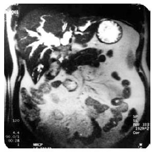
Figure 3 Magnetic resonance cholangiopancreaticography showing type 2 malignant hilarstricture.
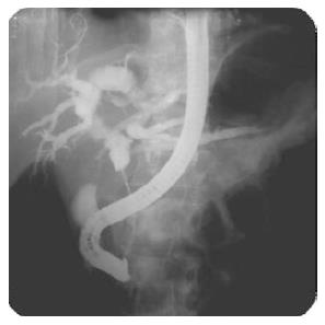
Figure 4 Type 1 malignant hilar stricture.
Figure 5 Type 2 malignant hilarstricture.
Figure 6 Type 2 malignant hilarstricture with bilateral guide wires.
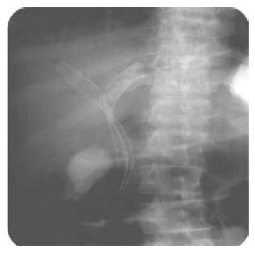
Figure 7 Bilateral metal stents in type 2 malignant hilar stricture.
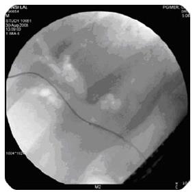
Figure 8 Air cholangiogram showing type 2 malignant hilar stricture.
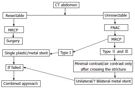
Figure 9 Approach to malignant hilar biliary strictures.
CT: Computed tomography; MRCP: Magnetic resonance cholangiopancreaticography; FNAC: Fine needle aspiration cytology.
- Citation: Singh RR, Singh V. Endoscopic management of hilar biliary strictures. World J Gastrointest Endosc 2015; 7(8): 806-813
- URL: https://www.wjgnet.com/1948-5190/full/v7/i8/806.htm
- DOI: https://dx.doi.org/10.4253/wjge.v7.i8.806


