Copyright
©The Author(s) 2015.
World J Gastrointest Endosc. Jun 25, 2015; 7(7): 670-674
Published online Jun 25, 2015. doi: 10.4253/wjge.v7.i7.670
Published online Jun 25, 2015. doi: 10.4253/wjge.v7.i7.670
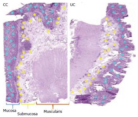
Figure 1 Illustrates histology-directed tissue compartment proteomics profiling using matrix-assisted-laser desorption/ionization mass spectrometry.
Digital photomicrographs acquired form histology and matrix-assisted-laser desorption/ionization sections are used to identify and designate sites of interest for profiling. Using bioinformatics technology comparisons are performed in both the training and independent test set samples between inflamed mucosa and inflamed submucosa Crohn’s colitis (CC) vs ulcerative colitis (UC). Tissue showing marked areas of pathological interest. Rings demonstrate matrix spots in mucosal (blue) and submucosal (yellow) layers (our unpublished data).
- Citation: Ballard BR, M’Koma AE. Gastrointestinal endoscopy biopsy derived proteomic patterns predict indeterminate colitis into ulcerative colitis and Crohn’s colitis. World J Gastrointest Endosc 2015; 7(7): 670-674
- URL: https://www.wjgnet.com/1948-5190/full/v7/i7/670.htm
- DOI: https://dx.doi.org/10.4253/wjge.v7.i7.670


