Copyright
©The Author(s) 2015.
World J Gastrointest Endosc. Jun 10, 2015; 7(6): 665-669
Published online Jun 10, 2015. doi: 10.4253/wjge.v7.i6.665
Published online Jun 10, 2015. doi: 10.4253/wjge.v7.i6.665
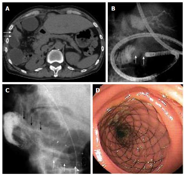
Figure 1 Metallic stent insertion with double-balloon endoscopy for malignant afferent loop obstruction in case 1.
A: Abdominal computed tomography showed the reconstructed jejunum that was expanded at the site of hepatectomy, expansion of intrahepatic bile ducts, and the stenosis of the reconstructed jejunum (arrows); B: The stenosis (arrows) was seen when the double-balloon endoscopy (DBE) reached the Roux-limb obstruction; C: An overtube was left to prevent bowel expansion. The DBE was then removed and an metallic stent (MS) (black arrows) was inserted through the overtube (white arrows) in combination with the over-the-wire technique; D: A Wallflex duodenal MS with a diameter of 2.2 cm and a length of 6.0 cm was deployed.
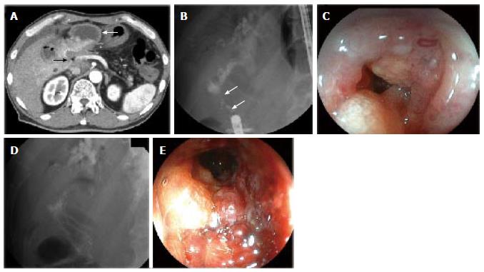
Figure 2 Metallic stent insertion with double-balloon endoscopy for malignant afferent loop obstruction in case 2.
A: Computed tomography showed ascites, dilation of the afferent loop (white arrow), and a surrounding soft density (black arrow); B: The double-balloon endoscopy reached the afferent loop obstruction (arrows); C: Stenosis with irregular mucosa was seen; D and E: A Niti-S duodenal metallic stent with a diameter of 2.2 cm and a length of 6.0 cm was inserted and deployed.
- Citation: Fujii M, Ishiyama S, Saito H, Ito M, Fujiwara A, Niguma T, Yoshioka M, Shiode J. Metallic stent insertion with double-balloon endoscopy for malignant afferent loop obstruction. World J Gastrointest Endosc 2015; 7(6): 665-669
- URL: https://www.wjgnet.com/1948-5190/full/v7/i6/665.htm
- DOI: https://dx.doi.org/10.4253/wjge.v7.i6.665


