Copyright
©The Author(s) 2015.
World J Gastrointest Endosc. Jun 10, 2015; 7(6): 617-627
Published online Jun 10, 2015. doi: 10.4253/wjge.v7.i6.617
Published online Jun 10, 2015. doi: 10.4253/wjge.v7.i6.617
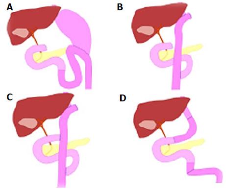
Figure 1 Schema of types of surgical anatomic reconstruction from gastrectomy.
A: Billroth II reconstruction; B: Roux-en-Y reconstruction; C: Double-tract reconstruction; D: Jejunal pouch interposition.
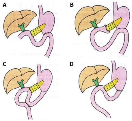
Figure 2 Schema of types of surgical anatomic reconstruction from pancreaticoduodenectomy.
A: The Whipple Method; B: The (modified) Child surgery; C: The Cattell Method; D: The Imanaga Method.
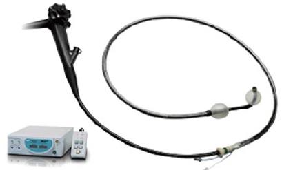
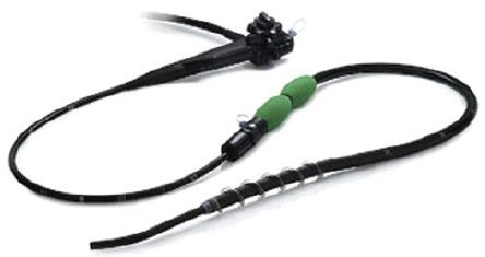
Figure 3 Double-balloon endoscopy.
The short type double balloon enteroscope (EC- 530B; FUJIFILM, Osaka, Japan) with a working channel of 2.8 mm diameter and a working length of 152 cm.
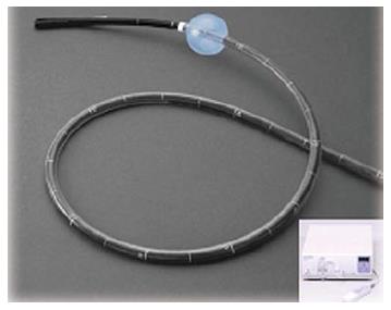
Figure 4 Single-balloon endoscopy.
The standard type double balloon enteroscope (SIF- Q260; Olympus Systems, Tokyo, Japan) with a working channel of 2.8 mm diameter and a working length of 200 cm.
Figure 5 Spiral endoscopy.
Discovery SB overtube over the snteroscope.
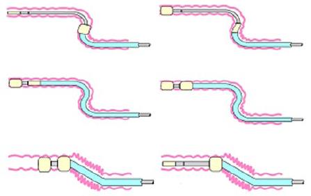
Figure 6 Schema of double-balloon endoscopy insertion.
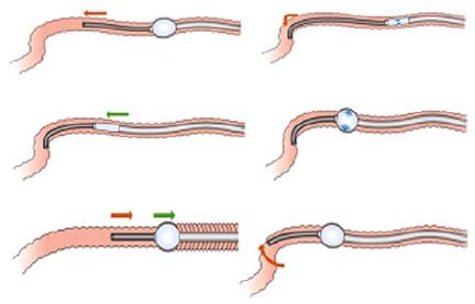
Figure 7 Schema of single-balloon endoscopy insertion.
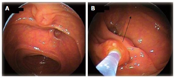
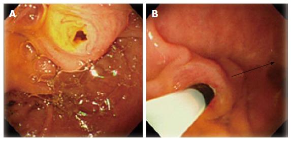
Figure 8 Biliary cannulation using double-balloon endoscopy in a patient with papilla.
A: Papilla when the blind end was accessed; B: Locating papilla in 6 o’clock direction in the monitor, and performing cannulation adjusting the axis of catheter into 12 o’clock direction along the biliary duct.
Figure 9 Biliary cannulation using single-balloon endoscopy in a patient with papilla.
A: Papilla when the blind end was accessed; B: Locating papilla in 8-9 o’clock direction in the monitor, and performing cannulation adjusting the axis of catheter into 3 o’clock direction along the biliary duct.
- Citation: Shimatani M, Takaoka M, Tokuhara M, Miyoshi H, Ikeura T, Okazaki K. Review of diagnostic and therapeutic endoscopic retrograde cholangiopancreatography using several endoscopic methods in patients with surgically altered gastrointestinal anatomy. World J Gastrointest Endosc 2015; 7(6): 617-627
- URL: https://www.wjgnet.com/1948-5190/full/v7/i6/617.htm
- DOI: https://dx.doi.org/10.4253/wjge.v7.i6.617


