Copyright
©The Author(s) 2015.
World J Gastrointest Endosc. Jun 10, 2015; 7(6): 582-592
Published online Jun 10, 2015. doi: 10.4253/wjge.v7.i6.582
Published online Jun 10, 2015. doi: 10.4253/wjge.v7.i6.582
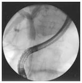
Figure 1 Distal cholangiocarcinoma during endoscopic retrograde cholangiography.
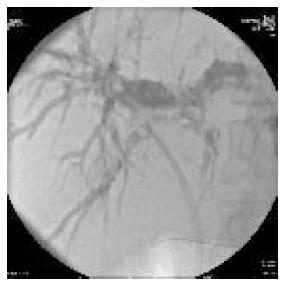
Figure 2 Endoscopic retrograde cholangiography with a plastic stent in the right hepatic duct.
However the left hepatic duct remains dilated.
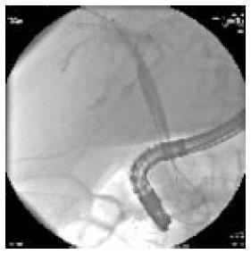
Figure 3 Use of covered self-expandable metal stent in patients with hylar cholangiocarcinoma.
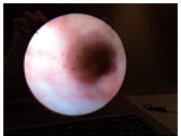
Figure 4 Visualization of biliary epithelium during SpyGlass.
- Citation: Bertani H, Frazzoni M, Mangiafico S, Caruso A, Manno M, Mirante VG, Pigò F, Barbera C, Manta R, Conigliaro R. Cholangiocarcinoma and malignant bile duct obstruction: A review of last decades advances in therapeutic endoscopy. World J Gastrointest Endosc 2015; 7(6): 582-592
- URL: https://www.wjgnet.com/1948-5190/full/v7/i6/582.htm
- DOI: https://dx.doi.org/10.4253/wjge.v7.i6.582


