Copyright
©The Author(s) 2015.
World J Gastrointest Endosc. Apr 16, 2015; 7(4): 370-380
Published online Apr 16, 2015. doi: 10.4253/wjge.v7.i4.370
Published online Apr 16, 2015. doi: 10.4253/wjge.v7.i4.370
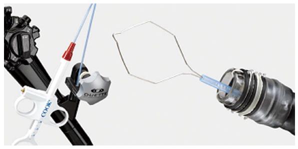
Figure 1 Multiband device (Duette).
A variceal ligation device is used to suck the lesion into the ligation cap, allowing it to be captured with a rubber band and resected with a hexagonal snare (Courtesy of Cook®).
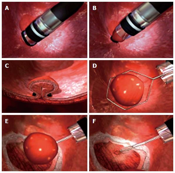
Figure 2 Multiband mucosectomy technical sequence (A to F) (Courtesy of Cook®).
A-C: Pseudopolyp that is created by suctioning the mucosa into the ligation cap and releasing a rubber band; D-F: Pseudopolyp resection by hexagonal snare.
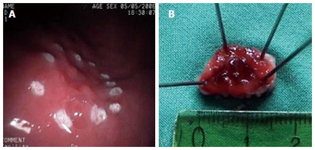
Figure 3 Early gastric cancer treated with multiband mucosectomy.
A: Argon plasma coagulation marks are placed 2-5 mm outside the margins of the lesion; B: Specimen resected (15 mm).
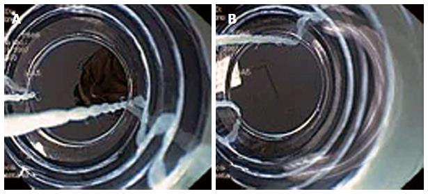
Figure 4 Best endoscopic views.
A: Wires positioned incorrectly; B: Wires positioned correctly (in line with the working cannel).
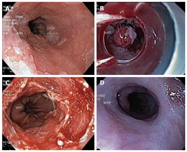
Figure 5 Stepwise radical multiband endoscopic resection of Barrett's esophagus with high-grade dysplasia (A to D).
A: A 3-cm long Barrett’s mucosa; B: Rubber band applied for resection; C: Circumferential resection; D: Complete neo-squamous re-epithelization.
Figure 6 Active bleeding post-multiband mucosectomy in Barrett's esophagus, effectively treated by adrenaline injection (A to C).
A: Active pumping bleeding; B: Adrenaline injection by needle; C: Cessation of bleeding.
- Citation: Espinel J, Pinedo E, Ojeda V, Rio MGD. Multiband mucosectomy for advanced dysplastic lesions in the upper digestive tract. World J Gastrointest Endosc 2015; 7(4): 370-380
- URL: https://www.wjgnet.com/1948-5190/full/v7/i4/370.htm
- DOI: https://dx.doi.org/10.4253/wjge.v7.i4.370


