Copyright
©The Author(s) 2015.
World J Gastrointest Endosc. Dec 25, 2015; 7(19): 1350-1354
Published online Dec 25, 2015. doi: 10.4253/wjge.v7.i19.1350
Published online Dec 25, 2015. doi: 10.4253/wjge.v7.i19.1350
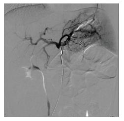
Figure 1 Computed tomography angiography with multiple serpiginous vessels in the left side of the bowel mesentery.
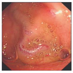
Figure 2 Suspicion of varices.
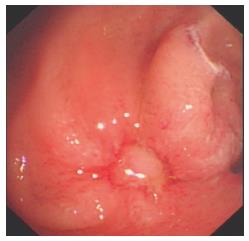
Figure 3 Appearance of jejunal varices using the double balloon enterosocpy.
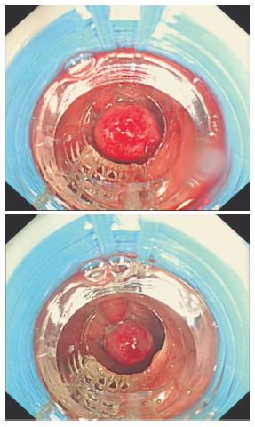
Figure 4 Banding of the jejunal varices using the operative gastroscope.
- Citation: Belsha D, Thomson M. Challenges of banding jejunal varices in an 8-year-old child. World J Gastrointest Endosc 2015; 7(19): 1350-1354
- URL: https://www.wjgnet.com/1948-5190/full/v7/i19/1350.htm
- DOI: https://dx.doi.org/10.4253/wjge.v7.i19.1350


