Copyright
©The Author(s) 2015.
World J Gastrointest Endosc. Dec 25, 2015; 7(19): 1311-1317
Published online Dec 25, 2015. doi: 10.4253/wjge.v7.i19.1311
Published online Dec 25, 2015. doi: 10.4253/wjge.v7.i19.1311
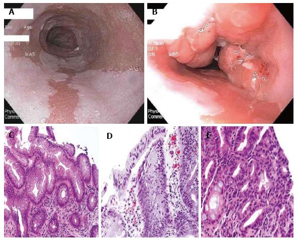
Figure 1 Histopathology pictures.
A: White-light endoscopic image of long segment BE; B: White-light endoscopic image of BE with nodular mucosa found to be HGD; C: Hematoxylin and eosin (HE) stain of Barrett’s mucosa; D: HE of Barrett’s mucosa with LGD; E: Barrett’s mucosa with HGD. Histopathology pictures courtesy of Purva Gopal, MD, Department of Pathology, University of Texas Southwestern Medical Center, Dallas, Texas. HGD: High-grade dysplasia; LGD: Low-grade dysplasia; BE: Barrett’s esophagus.
- Citation: Vance RB, Dunbar KB. Endoscopic options for treatment of dysplasia in Barrett’s esophagus. World J Gastrointest Endosc 2015; 7(19): 1311-1317
- URL: https://www.wjgnet.com/1948-5190/full/v7/i19/1311.htm
- DOI: https://dx.doi.org/10.4253/wjge.v7.i19.1311


