Copyright
©The Author(s) 2015.
World J Gastrointest Endosc. Aug 25, 2015; 7(11): 1014-1022
Published online Aug 25, 2015. doi: 10.4253/wjge.v7.i11.1014
Published online Aug 25, 2015. doi: 10.4253/wjge.v7.i11.1014
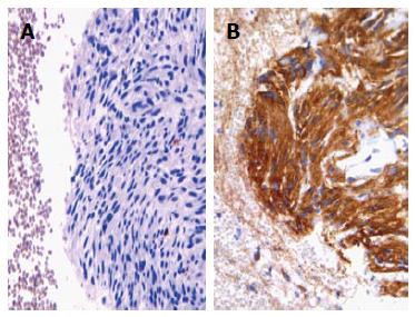
Figure 1 Cell block from gastrointestinal stromal tumour exhibiting aggregates of spindle cells with elongated nuclei (haematoxylin-eosin, × 200) (A), with an evident immunoreactivity for CD117 (immunoperoxidase, × 200, Mayer’s Haemalum nuclear counterstain) (B).
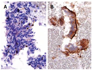
Figure 2 Spindle cells of gastric gastrointestinal stromal tumour documented only a sporadic nuclear Ki67 immunopositivity (immunoperoxidase, × 400, Mayer’s Haemalum nuclear counterstain) (A) in benign contaminant gastrointestinal cells, the apical cytoplasm showed a peculiar CD10 immunoreactivity (immunoperoxidase, × 400, Mayer’s Haemalum nuclear counterstain) (B).
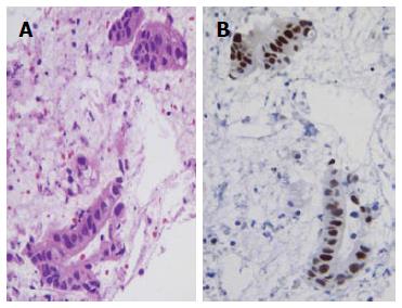
Figure 3 Well differentiated pancreatic carcinoma with a pseudo-glandular pattern (haematoxylin-eosin, × 400) (A), a nuclear strong p53 immunostaining was encountered in neoplastic elements (immunoperoxidase, × 400, Mayer’s Haemalum nuclear counterstain) (B).
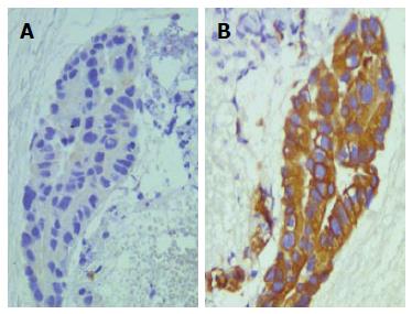
Figure 4 Peripheral cholangiocarcinoma with a papillary pattern (haematoxylin-eosin, × 400) (A), in a serial section obtained from CBP neoplastic elements exhibited an evident cytokeratin 7 immunoreaction (B) (immunoperoxidase, × 400, Mayer’s Haemalum nuclear counterstain).
- Citation: Ieni A, Barresi V, Todaro P, Caruso RA, Tuccari G. Cell-block procedure in endoscopic ultrasound-guided-fine-needle-aspiration of gastrointestinal solid neoplastic lesions. World J Gastrointest Endosc 2015; 7(11): 1014-1022
- URL: https://www.wjgnet.com/1948-5190/full/v7/i11/1014.htm
- DOI: https://dx.doi.org/10.4253/wjge.v7.i11.1014


