Copyright
©The Author(s) 2015.
World J Gastrointest Endosc. Aug 10, 2015; 7(10): 987-994
Published online Aug 10, 2015. doi: 10.4253/wjge.v7.i10.987
Published online Aug 10, 2015. doi: 10.4253/wjge.v7.i10.987
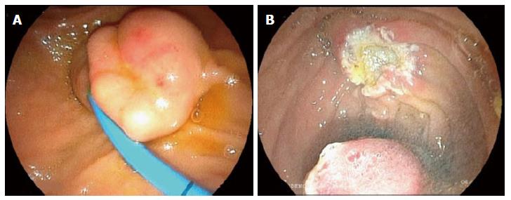
Figure 1 Endoscopic view of neuroendocrine tumor of the papilla with Fujinon intelligent chromo endoscopy.
A: This picture show the depression in the center of the lesion; B: The picture shows the aspect of the papillary region after the “en bloc” resection.
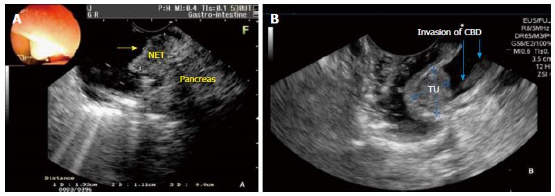
Figure 2 Endoscopic ultrasound staging of the duodenal papilla.
A: Patient of the Figure 1. Endoscopic ultrasound staging shows the regular and hypoechoic nodule (1.93 cm) in the papilla without infiltration of the duodenal wall and pancreatic gland. The staging was uT1N0Mx; B: This picture shows the papillary tumor with 1.72 cm with invasion of the common bile duct wall (blue arrows). NET: Neuroendocrine tumor; TU: Tumor; CBD: Common bile duct.
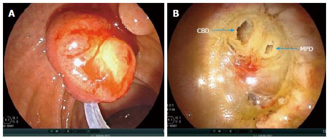
Figure 3 Endoscopic papillectomy immediately after endoscopic ultrasound for staging.
A: En bloc resection of the tumor, after the snare is widely opened, duodenoscope is pushed in a craniocaudal direction; B: The endoscopic view of the common bile duct and main pancreatic duct (blue arrows) after a complete en bloc resection of the papillary tumor. CBD: Common bile duct; MPD: Main pancreatic duct.
- Citation: Ardengh JC, Kemp R, Lima-Filho &R, Santos JSD. Endoscopic papillectomy: The limits of the indication, technique and results. World J Gastrointest Endosc 2015; 7(10): 987-994
- URL: https://www.wjgnet.com/1948-5190/full/v7/i10/987.htm
- DOI: https://dx.doi.org/10.4253/wjge.v7.i10.987


