Copyright
©The Author(s) 2015.
World J Gastrointest Endosc. Aug 10, 2015; 7(10): 950-959
Published online Aug 10, 2015. doi: 10.4253/wjge.v7.i10.950
Published online Aug 10, 2015. doi: 10.4253/wjge.v7.i10.950
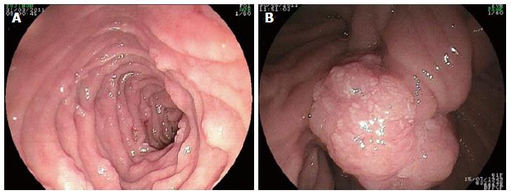
Figure 1 Endoscopic view showing a stage II disease (10-20 small duodenal adenomas with tubular histology) in A, and a large papilla lesion which biopsy revealed a well-moderated carcinoma in B.
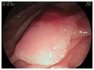
Figure 2 Detection of a prominent papilla of Vater with a solitary adenoma with the help of a side-viewing endoscope.
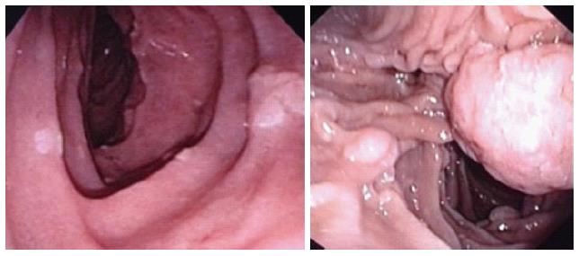
Figure 3 Endoscopic aspect of a stage I patient exhibiting 3 adenomatous-tubular polyps with low-grade dysplasia (left); on the right, one may observe a 6 mm tubulo-villous polyp with severe dysplasia, along with other smaller adenomas diagnosed in another patient (stage IV disease).
- Citation: Campos FG, Sulbaran M, Safatle-Ribeiro AV, Martinez CAR. Duodenal adenoma surveillance in patients with familial adenomatous polyposis. World J Gastrointest Endosc 2015; 7(10): 950-959
- URL: https://www.wjgnet.com/1948-5190/full/v7/i10/950.htm
- DOI: https://dx.doi.org/10.4253/wjge.v7.i10.950


