Copyright
©2014 Baishideng Publishing Group Inc.
World J Gastrointest Endosc. Jul 16, 2014; 6(7): 328-333
Published online Jul 16, 2014. doi: 10.4253/wjge.v6.i7.328
Published online Jul 16, 2014. doi: 10.4253/wjge.v6.i7.328
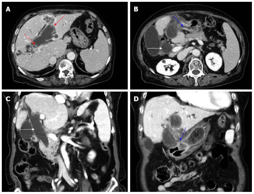
Figure 1 Computed tomography of the abdomen showed markedly dilated common bile duct (white arrows) and left intrahepatic duct with left intrahepatic duct penetrating into the antrum of stomach and fistula formation (blue arrows) and papillary projections along the dilated bile duct (red arrows) and no definite visible mass in the left intrahepatic duct (A-D).
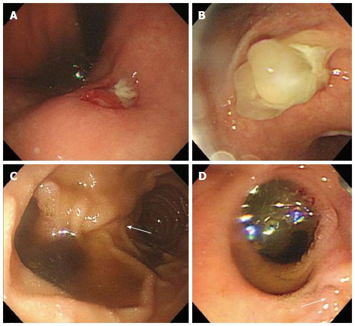
Figure 2 Endoscopic finding showed a round ulcerated lesion (A) and extruding white thick mucin from the opening at the lesser curvature of the antrum during endoscopic suction (B) and another wide opening of the fistula with mucin excretion proximal to the original papillary orifice (white arrows) (C, D).
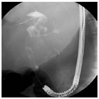
Figure 3 Endoscopic retrograde cholangiopancreatography finding through the choledochoduodenal fistula near the papillary orifice showed moderately to severely dilated common bile duct and proximal left intrahepatic duct with amorphous, partial intraluminal filling of the contrast.
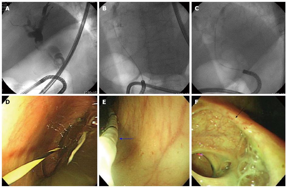
Figure 4 Cholangiogram using ultra-slim endoscope showed moderately to severely dilated common bile duct and both proximal intrahepatic duct with the amorphous, partial intraluminal filling in the bile duct (A-C) and the suction of thick mucus after advancement into bile duct and approach up to the cystic duct (black arrow) level using anchoring of the balloon catheter (blue arrow) was ineffective (D-F).
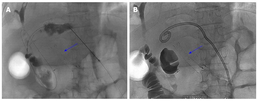
Figure 5 Cholangiogram obtained after contrast injection through the access needle into the left intrahepatic duct showed moderately to severely dilated left intrahepatic duct and common bile duct with the amorphous intraluminal filling consistent with the endoscopic retrograde cholangiopancreatography finding and the metal stent (blue arrow) previously inserted into the choledochogastric fistula for facilitating endoscopic suction of mucin through the stent was in place (A, B).
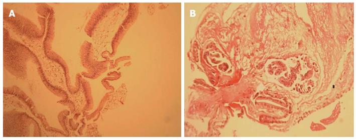
Figure 6 Pathology of the specimen from the proximal portion of the choledochogastric fistula showed a low grade adenoma [hematoxylin and eosin stain, × 200 (left), × 400 (right)].
- Citation: Hong MY, Yu DW, Hong SG. Intraductal papillary mucinous neoplasm of the bile duct with gastric and duodenal fistulas. World J Gastrointest Endosc 2014; 6(7): 328-333
- URL: https://www.wjgnet.com/1948-5190/full/v6/i7/328.htm
- DOI: https://dx.doi.org/10.4253/wjge.v6.i7.328


