Copyright
©2014 Baishideng Publishing Group Inc.
World J Gastrointest Endosc. Jun 16, 2014; 6(6): 260-265
Published online Jun 16, 2014. doi: 10.4253/wjge.v6.i6.260
Published online Jun 16, 2014. doi: 10.4253/wjge.v6.i6.260
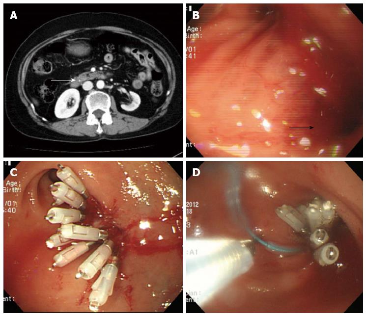
Figure 1 Initial abdominal computed tomography and endoscopic findings during endoscopic retrograde cholangiopancreatography.
A: Computed tomography showed a small common bile duct stone (thin white arrow); B: During endoscopic retrograde cholangiopancreatography, a 10 mm-sized perforation developed in the lateral wall of the duodenal bulb during inadvertent rapid withdrawal of the duodenoscope (thick black arrow); C and D: Multiple hemoclips and an endoloop were immediately applied for the defect closure.
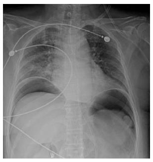
Figure 2 Chest X-ray after endoscopic retrograde cholangiopancreatography.
The scan shows a large amount of pneumoperitoneum below both the hemidiaphragms.
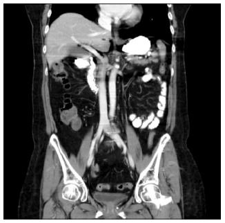
Figure 3 Follow-up abdominal computed tomography after duodenal perforation.
No contrast leakage was detected into the peritoneum at the periduodenal lesion after endoscopic treatment; however, a moderate amount of pneumoperitoneum was present.
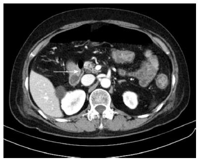
Figure 4 Second follow-up abdominal computed tomography after duodenal perforation.
The scan shows a small fistula communicating with the peritoneal space, at the previous perforation site in the duodenal bulb (arrow), and absence of a common bile duct stone.
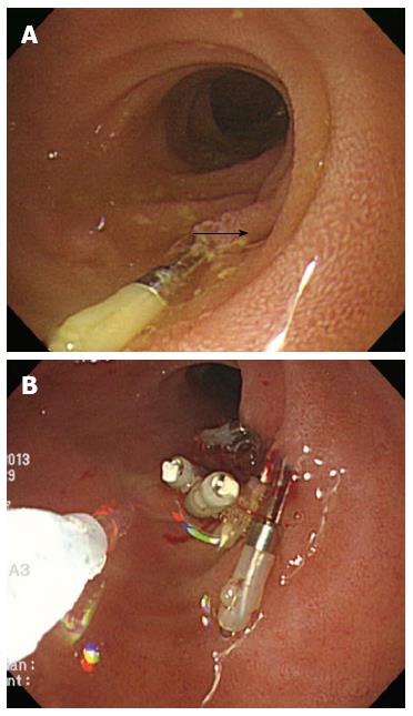
Figure 5 Endoscopic findings of the second endoscopic closure.
A: A suspicious fistulous opening was detected at the previous perforation site (arrow); B: An application of additional multiple hemoclips and fibrin glue injection was successfully performed at the site of the suspicious fistulous opening.
- Citation: Yu DW, Hong MY, Hong SG. Endoscopic treatment of duodenal fistula after incomplete closure of ERCP-related duodenal perforation. World J Gastrointest Endosc 2014; 6(6): 260-265
- URL: https://www.wjgnet.com/1948-5190/full/v6/i6/260.htm
- DOI: https://dx.doi.org/10.4253/wjge.v6.i6.260


