Copyright
©2014 Baishideng Publishing Group Inc.
World J Gastrointest Endosc. Dec 16, 2014; 6(12): 625-629
Published online Dec 16, 2014. doi: 10.4253/wjge.v6.i12.625
Published online Dec 16, 2014. doi: 10.4253/wjge.v6.i12.625
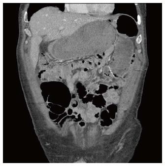
Figure 1 Computed tomography scan showed a fluid-filled gastric remnant, a wider duodenal wall, and multiple fluid levels through proximal small intestine.
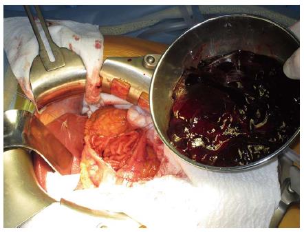
Figure 2 Blood evacuated from gastric remnant.
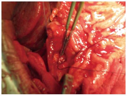
Figure 3 Thirty millimeters wide ulcer on the posterior part of the second portion of the duodenum with a bleeding branch of gastro-duodenal artery marked with tweezers.
- Citation: Ivanecz A, Sremec M, Ćeranić D, Potrč S, Skok P. Life threatening bleeding from duodenal ulcer after Roux-en-Y gastric bypass: Case report and review of the literature. World J Gastrointest Endosc 2014; 6(12): 625-629
- URL: https://www.wjgnet.com/1948-5190/full/v6/i12/625.htm
- DOI: https://dx.doi.org/10.4253/wjge.v6.i12.625


