Copyright
©2014 Baishideng Publishing Group Inc.
World J Gastrointest Endosc. Nov 16, 2014; 6(11): 525-533
Published online Nov 16, 2014. doi: 10.4253/wjge.v6.i11.525
Published online Nov 16, 2014. doi: 10.4253/wjge.v6.i11.525
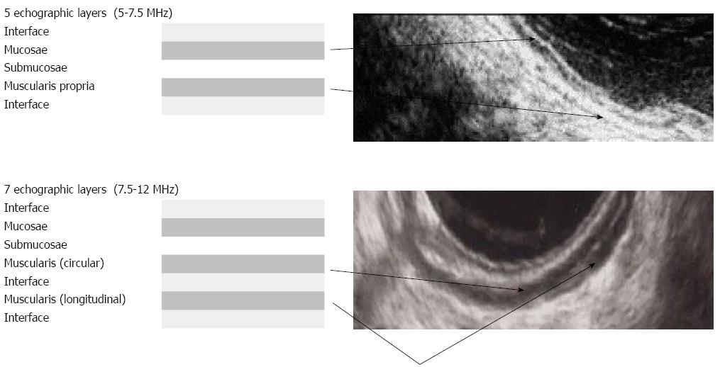
Figure 1 Digestive walls echoic stratifications depending on frequencies.
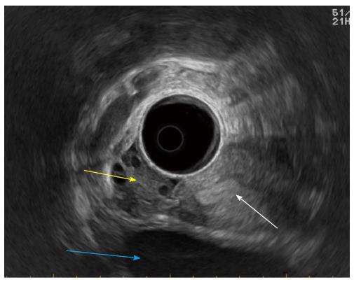
Figure 2 Normal peri-rectal anatomy.
White arrow: Uterus; Yellow arrow: Right ovary; Blue arrow: Bladder.
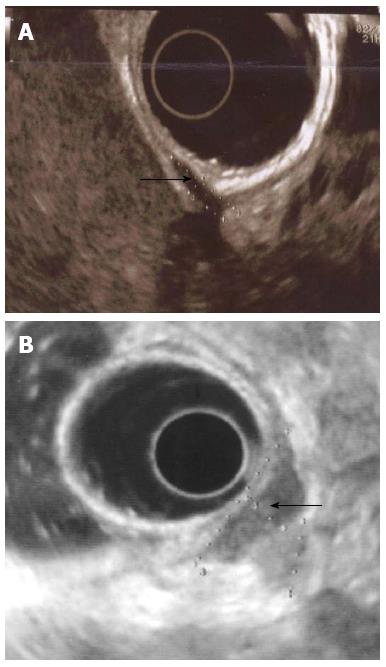
Figure 3 Different types of muscularis propria infiltration.
A: Infiltration limited to external muscularis propria; B: Infiltration of the entire thickness of the muscularis propria.
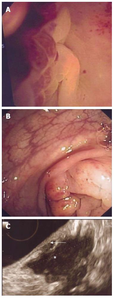
Figure 4 Endoscopic and echographic presentations of recto-sigmoid endometriosis.
A: Mucosae infiltration; B: Sigmoïd stenosis; C: Sub-mucosae (white arrow) and muscularis (white star) infiltrations.
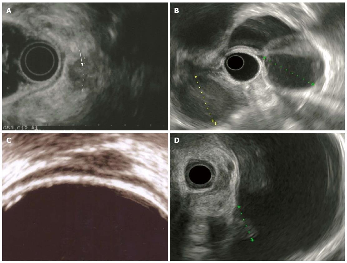
Figure 5 Other pelvic endometriosis locations.
A: Infiltrated Torus; B: Bilateral Ovarian endometriomas; C: Infiltrated utero-sacral ligament; D: Bladder nodule.
- Citation: Roseau G. Recto-sigmoid endoscopic-ultrasonography in the staging of deep infiltrating endometriosis. World J Gastrointest Endosc 2014; 6(11): 525-533
- URL: https://www.wjgnet.com/1948-5190/full/v6/i11/525.htm
- DOI: https://dx.doi.org/10.4253/wjge.v6.i11.525


