Copyright
©2014 Baishideng Publishing Group Inc.
World J Gastrointest Endosc. Oct 16, 2014; 6(10): 488-492
Published online Oct 16, 2014. doi: 10.4253/wjge.v6.i10.488
Published online Oct 16, 2014. doi: 10.4253/wjge.v6.i10.488
Figure 1 Endoscopic views of the vascular ectasia.
A: Bleeding at the horizontal portion of the duodenum; B: View through the band-ligator device; C: Endoscopic band ligation was performed.
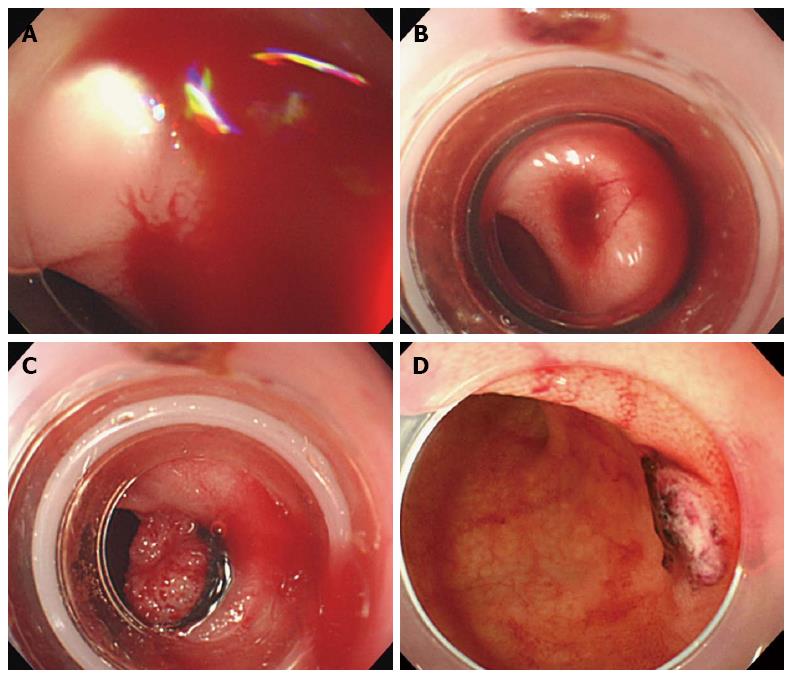
Figure 2 Endoscopic views of the ileal diverticulum.
A: Active bleeding; B: View through the band-ligator device; C: Endoscopic band ligation was performed. However, eversion of the banded diverticulum was not sufficient; D: Repeat endoscopic view of O-band dislodgement and ulcer formation at the banded site.
- Citation: Ikeya T, Ishii N, Shimamura Y, Nakano K, Ego M, Nakamura K, Takagi K, Fukuda K, Fujita Y. Endoscopic band ligation for bleeding lesions in the small bowel. World J Gastrointest Endosc 2014; 6(10): 488-492
- URL: https://www.wjgnet.com/1948-5190/full/v6/i10/488.htm
- DOI: https://dx.doi.org/10.4253/wjge.v6.i10.488


