Copyright
©2013 Baishideng Publishing Group Co.
World J Gastrointest Endosc. Sep 16, 2013; 5(9): 450-454
Published online Sep 16, 2013. doi: 10.4253/wjge.v5.i9.450
Published online Sep 16, 2013. doi: 10.4253/wjge.v5.i9.450
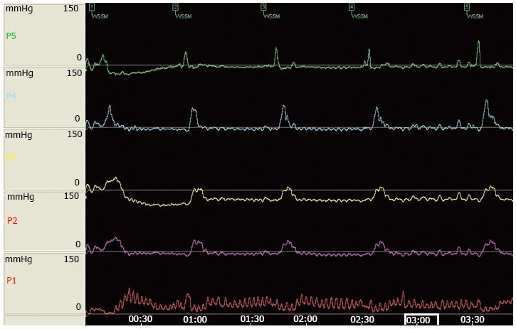
Figure 1 Esophageal manometric findings of elevated intra-esophageal resting pressure > 4 mmHg, localized at 36 cm from the nose, with superimposed cyclic pressure spikes with a frequency of 60-100/min with absence of relaxation in response to swallow (see P1 in the second swallow), typical of esophageal vascular compression.
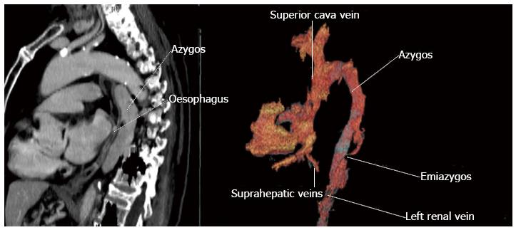
Figure 2 Computed tomography view of a dilated ascending thoracic aorta with no presence of the inferior vena cava with azygos continuation, cause of the vascular compression of esophagus.
- Citation: Campo SMA, Zullo A, Scandavini CM, Frezza B, Cerro P, Balducci G. Pseudoachalasia: A peculiar case report and review of the literature. World J Gastrointest Endosc 2013; 5(9): 450-454
- URL: https://www.wjgnet.com/1948-5190/full/v5/i9/450.htm
- DOI: https://dx.doi.org/10.4253/wjge.v5.i9.450


