Copyright
©2013 Baishideng Publishing Group Co.
World J Gastrointest Endosc. Jun 16, 2013; 5(6): 297-299
Published online Jun 16, 2013. doi: 10.4253/wjge.v5.i6.297
Published online Jun 16, 2013. doi: 10.4253/wjge.v5.i6.297
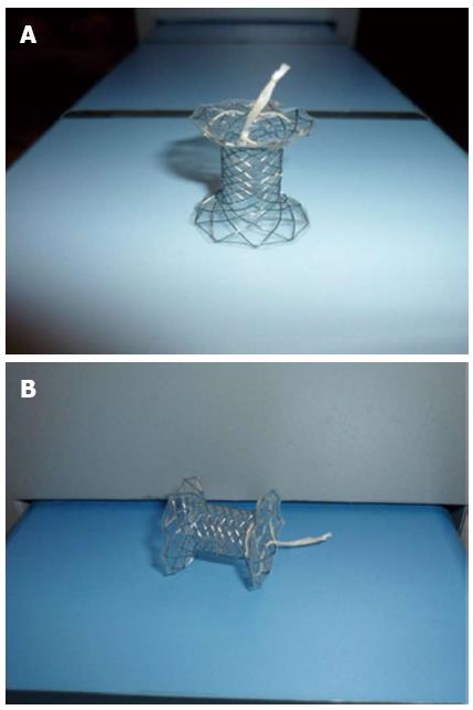
Figure 1 Novel “NAGI” covered self-expanding metallic stents with a 10 mm center and 20 mm ends (A and B).
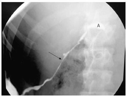
Figure 2 Presence of stenosis (arrow) and leak (A) of the main pancreatic duct.
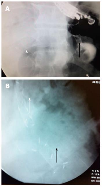
Figure 3 Fluoroscopy image at basal (A) and at after 6 mo (B) of follow-up: Biliary stent (white arrows) and Nagi stent through cystogastrostomy (black arrows).
- Citation: Téllez-Ávila FI, Villalobos-Garita &, Ramírez-Luna M&. Use of a novel covered self-expandable metal stent with an anti-migration system for endoscopic ultrasound-guided drainage of a pseudocyst. World J Gastrointest Endosc 2013; 5(6): 297-299
- URL: https://www.wjgnet.com/1948-5190/full/v5/i6/297.htm
- DOI: https://dx.doi.org/10.4253/wjge.v5.i6.297


