Copyright
©2013 Baishideng Publishing Group Co.
World J Gastrointest Endosc. May 16, 2013; 5(5): 270-272
Published online May 16, 2013. doi: 10.4253/wjge.v5.i5.270
Published online May 16, 2013. doi: 10.4253/wjge.v5.i5.270
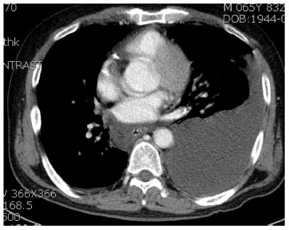
Figure 1 Chest computed tomography shows a left pleural effusion and peri-esophageal fluid collection.
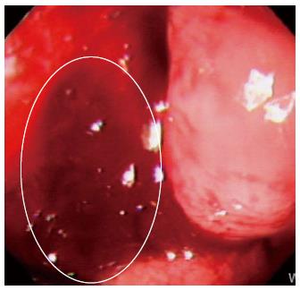
Figure 2 Upper endoscopy shows a 15 mm × 12 mm perforation with stigmata of recent bleeding distal to the Z-line on the left side of the esophagus.
- Citation: Yu JY, Kim SK, Jang EC, Yeom JO, Kim SY, Cho YS. Boerhaave’s syndrome during bowel preparation with polyethylene glycol in a patient with postpolypectomy bleeding. World J Gastrointest Endosc 2013; 5(5): 270-272
- URL: https://www.wjgnet.com/1948-5190/full/v5/i5/270.htm
- DOI: https://dx.doi.org/10.4253/wjge.v5.i5.270


