Copyright
©2013 Baishideng Publishing Group Co.
World J Gastrointest Endosc. May 16, 2013; 5(5): 246-250
Published online May 16, 2013. doi: 10.4253/wjge.v5.i5.246
Published online May 16, 2013. doi: 10.4253/wjge.v5.i5.246
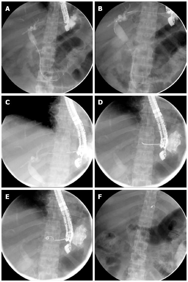
Figure 1 Endoscopic ultrasound-guided biliary drainage followed by endocoil placement.
A,B: After successful endoscopic ultrasound cannulation of segment 3 duct a guide wire is passed into the duodenum, the stricture dilated and a 10 mm × 80 mm uncovered biliary stent deployed; C: Next the catheter is withdrawn with the guide wire still in place; D, E: Finally the guide wire is removed with the catheter in position in the track between the dilated segment and the liver capsule an endocoil is advanced and deployed using a standard 0.035 guide wire; F: The final result is shown after stenting of the stricture in the duodenum, showing the biliary stent, duodenal stent and endocoil.
- Citation: van der Merwe SW, Omoshoro-Jones J, Sanyika C. Endocoil placement after endoscopic ultrasound-guided biliary drainage may prevent a bile leak. World J Gastrointest Endosc 2013; 5(5): 246-250
- URL: https://www.wjgnet.com/1948-5190/full/v5/i5/246.htm
- DOI: https://dx.doi.org/10.4253/wjge.v5.i5.246


