Copyright
©2013 Baishideng Publishing Group Co.
World J Gastrointest Endosc. May 16, 2013; 5(5): 226-230
Published online May 16, 2013. doi: 10.4253/wjge.v5.i5.226
Published online May 16, 2013. doi: 10.4253/wjge.v5.i5.226
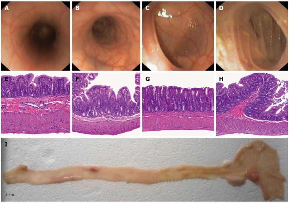
Figure 1 Representative pictures of in vivo colonoscopy (A-D), photomicrograph of histological study with hematoxylin and eosin stains of colon sections (E-H) in rats at 3 (A,E), 7 (B,F), 14 (C,G) and 20 cm (D, H) from the anal margin.
Macroscopic picture of the entire colon (I).
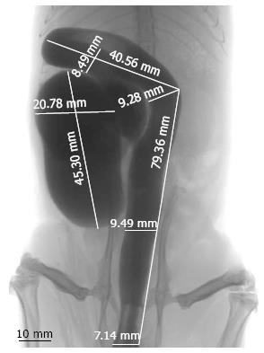
Figure 2 Tomographic picture of rat colon with length and diameter measurements.
- Citation: Bartolí R, Boix J, Òdena G, De la Ossa ND, de Vega VM, Lorenzo-Zúñiga V. Colonoscopy in rats: An endoscopic, histological and tomographic study. World J Gastrointest Endosc 2013; 5(5): 226-230
- URL: https://www.wjgnet.com/1948-5190/full/v5/i5/226.htm
- DOI: https://dx.doi.org/10.4253/wjge.v5.i5.226


