Copyright
©2013 Baishideng Publishing Group Co.
World J Gastrointest Endosc. Apr 16, 2013; 5(4): 197-200
Published online Apr 16, 2013. doi: 10.4253/wjge.v5.i4.197
Published online Apr 16, 2013. doi: 10.4253/wjge.v5.i4.197
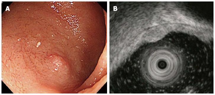
Figure 1 Endoscopic and endoscopic ultrasonography findings.
A: Endoscopic image showing an elevated lesion in the anterior wall of duodenal bulb; B: Endoscopic ultrasonography image of the lesion, a 3 mm hypoechoic mass lesion that was located in the submucosal layer.
Figure 2 Endoscopic image showing the endoscopic mucosal resection with circumferential mucosal incision procedures.
A-C: The entire lesion was removed en bloc.
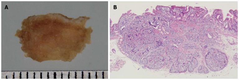
Figure 3 Histopathologic assessment of the resected specimen.
A: Macroscopic view of the resected specimen; B: Well-differentiated neuroendocrine tumor was confined to the submucosa (hematoxylin and eosin, original magnification × 20).
- Citation: Otaki Y, Homma K, Nawata Y, Imaizumi K, Arai S. Endoscopic mucosal resection with circumferential mucosal incision of duodenal carcinoid tumors. World J Gastrointest Endosc 2013; 5(4): 197-200
- URL: https://www.wjgnet.com/1948-5190/full/v5/i4/197.htm
- DOI: https://dx.doi.org/10.4253/wjge.v5.i4.197


