Copyright
©2013 Baishideng Publishing Group Co.
World J Gastrointest Endosc. Jan 16, 2013; 5(1): 6-13
Published online Jan 16, 2013. doi: 10.4253/wjge.v5.i1.6
Published online Jan 16, 2013. doi: 10.4253/wjge.v5.i1.6
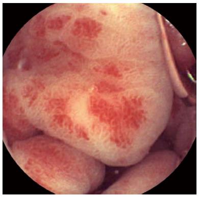
Figure 1 Endoscopic appearance of gastric antral vascular ectasia: Red spots radially departing from pylorus and involving the gastric antrum.
Figure 2 Videocapsule image of gastric antral vascular ectasia.
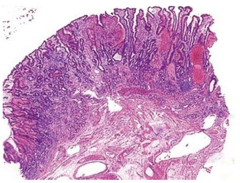
Figure 3 Gastric biopsy showing prominent vascular congestion with thrombosis of the vasculature.
The surrounding glands appear regenerative and the vessels in the submucosa are dilated and sclerotic.
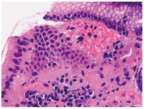
Figure 4 Higher magnification of one of the thrombosed vessels.
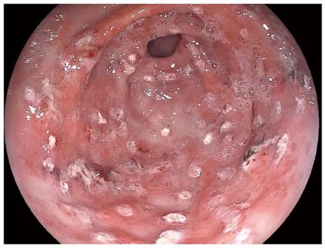
Figure 5 Argon plasma coagulation treatment of gastric antral vascular ectasia in patient with transfusion-dependent anaemia.
- Citation: Fuccio L, Mussetto A, Laterza L, Eusebi LH, Bazzoli F. Diagnosis and management of gastric antral vascular ectasia. World J Gastrointest Endosc 2013; 5(1): 6-13
- URL: https://www.wjgnet.com/1948-5190/full/v5/i1/6.htm
- DOI: https://dx.doi.org/10.4253/wjge.v5.i1.6


