Copyright
©2013 Baishideng Publishing Group Co.
World J Gastrointest Endosc. Jan 16, 2013; 5(1): 34-38
Published online Jan 16, 2013. doi: 10.4253/wjge.v5.i1.34
Published online Jan 16, 2013. doi: 10.4253/wjge.v5.i1.34
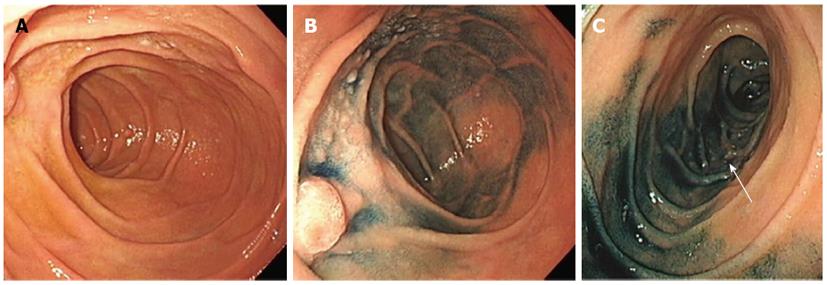
Figure 1 Images obtained during esophagogastroduodenoscopy.
A: Mormal white-light observation revealed whitish polyps around the ampulla of Vater; B: Indigo carmine spraying increased the contrast of the lesion; C: Whitish polyps were also seen in the third portion (arrow).
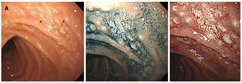
Figure 2 Close-up observation of the follicular lymphoma lesion.
A: Enlarged whitish villi (arrowhead) and tiny submucosal whitish depositions (arrow); B: Indigo carmine spraying visualized these microsurface structures more clearly; C: Narrow-band imaging view also emphasized microsurface structures.
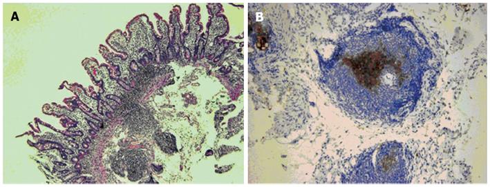
Figure 3 Pathological evaluation of the biopsy samples.
A: Monotonous proliferation of small- to medium-sized lymphoid cells which formed lymphoid follicles and infiltrated into the villi; B: The lymphoma cells were positive for CD10 expression.
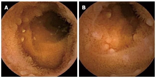
Figure 4 Multiple jejunal involvement revealed by video capsule endoscopy.
- Citation: Iwamuro M, Kawai Y, Takata K, Kawano S, Yoshino T, Okada H, Yamamoto K. Primary intestinal follicular lymphoma: How to identify follicular lymphoma by routine endoscopy. World J Gastrointest Endosc 2013; 5(1): 34-38
- URL: https://www.wjgnet.com/1948-5190/full/v5/i1/34.htm
- DOI: https://dx.doi.org/10.4253/wjge.v5.i1.34


