Copyright
©2013 Baishideng Publishing Group Co.
World J Gastrointest Endosc. Jan 16, 2013; 5(1): 29-33
Published online Jan 16, 2013. doi: 10.4253/wjge.v5.i1.29
Published online Jan 16, 2013. doi: 10.4253/wjge.v5.i1.29
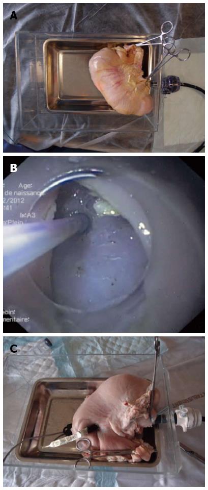
Figure 1 Endoscope.
A: The “in vitro” pig stomach model with the endoscope in place; B: View over muscularis propria from the gastric lumen in the submucosal space created by endoscopic submucosal dissection. Muscularis propria is about to be punctured (“peritoneoscopy”); C: Endoscope outside of the stomach simulating peritoneoscopy, with the dilated balloon in the working channel.
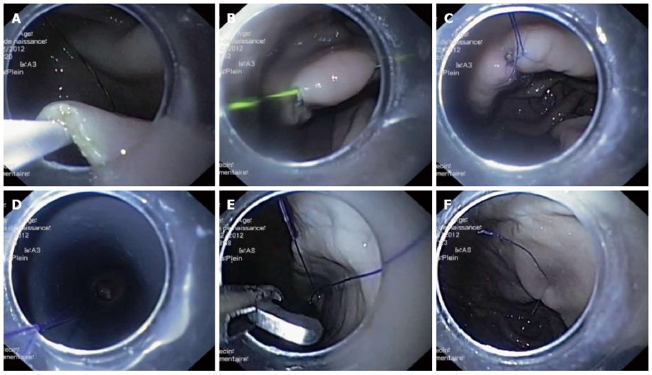
Figure 2 Natural orifice translumenal endoscopic surgery.
A: Puncture of the mucosa from the submucosal space on the right side of the incision and passing a guidewire into the gastric lumen; B: The guidewire traverses the mucosa on the right side of the incision, both ends are outside; C: Surgical wire replaces the guidewire first on the right side, then is passed through both sides after replacing the guidewire on the left side; D: A loop formed outside is pushed with the endoscope (here at the rim of the transparent cap) at the mucosal incision so as to tighten the knot; E Cutting the wire ends; F: The final aspect of the surgical knot.
-
Citation: Ciocirlan M, Ionescu ME, Diculescu MM. Endoscopic knot tying:
In vitro assessment in a porcine stomach model. World J Gastrointest Endosc 2013; 5(1): 29-33 - URL: https://www.wjgnet.com/1948-5190/full/v5/i1/29.htm
- DOI: https://dx.doi.org/10.4253/wjge.v5.i1.29


