Copyright
©2012 Baishideng.
World J Gastrointest Endosc. Sep 16, 2012; 4(9): 432-433
Published online Sep 16, 2012. doi: 10.4253/wjge.v4.i9.432
Published online Sep 16, 2012. doi: 10.4253/wjge.v4.i9.432
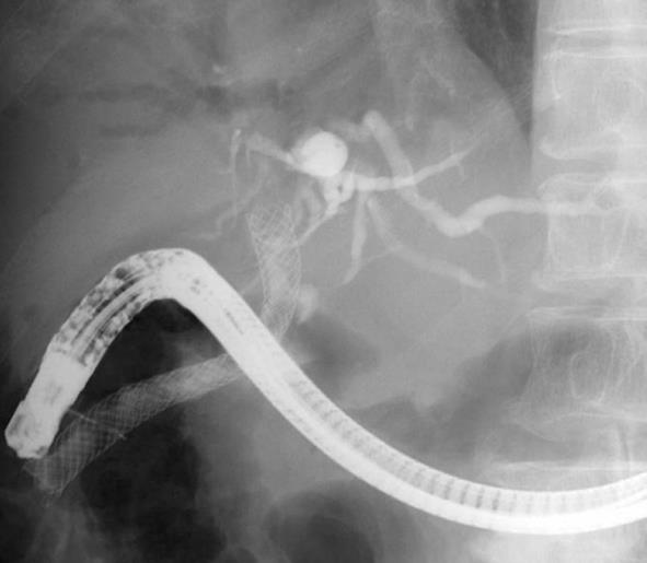
Figure 1 Fluoroscopic image a showing metal stent grasped with a snare.
It was impossible to remove the stent.
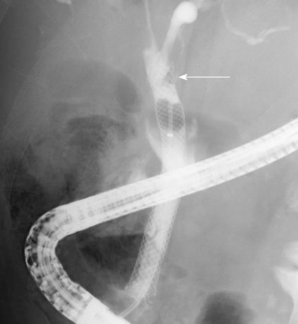
Figure 2 Fluoroscopic image showing a filling defect at the proximal end of a partially covered Wallflex stent (arrow).
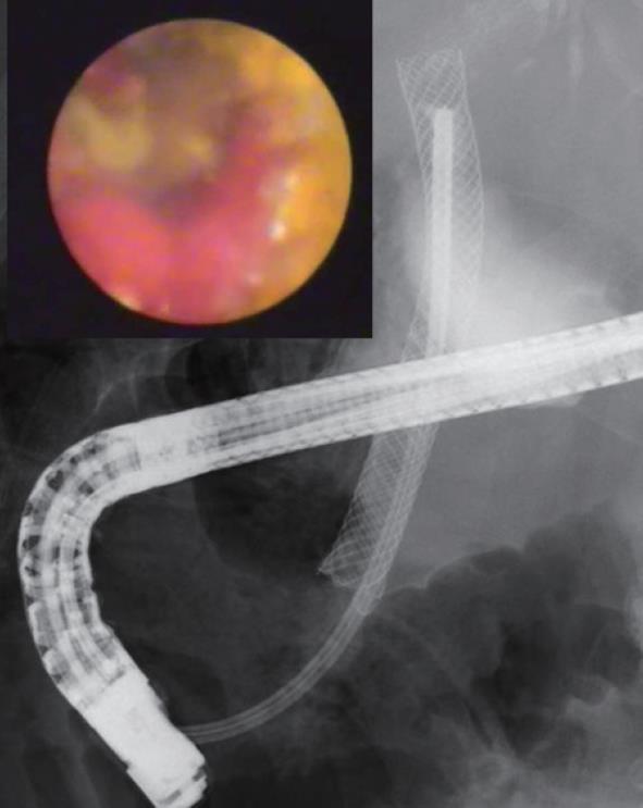
Figure 3 SpyGlass cholangioscopic image showing mucosal hyperplasia in an uncovered portion of a partially covered Wallflex stent.
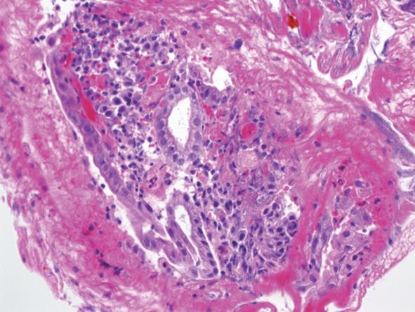
Figure 4 Biopsy specimen of mucosal hyperplasia showing inflammatory bile duct mucosa without malignancy.
- Citation: Kawakubo K, Isayama H, Sasahira N, Kogure H, Miyabayashi K, Hirano K, Tada M, Koike K. Mucosal hyperplasia in an uncovered portion of partially covered metal stent. World J Gastrointest Endosc 2012; 4(9): 432-433
- URL: https://www.wjgnet.com/1948-5190/full/v4/i9/432.htm
- DOI: https://dx.doi.org/10.4253/wjge.v4.i9.432


