Copyright
©2012 Baishideng.
World J Gastrointest Endosc. Sep 16, 2012; 4(9): 398-404
Published online Sep 16, 2012. doi: 10.4253/wjge.v4.i9.398
Published online Sep 16, 2012. doi: 10.4253/wjge.v4.i9.398
Figure 1 Needle-knife fistulotomy in Billroth II and removal of stone.
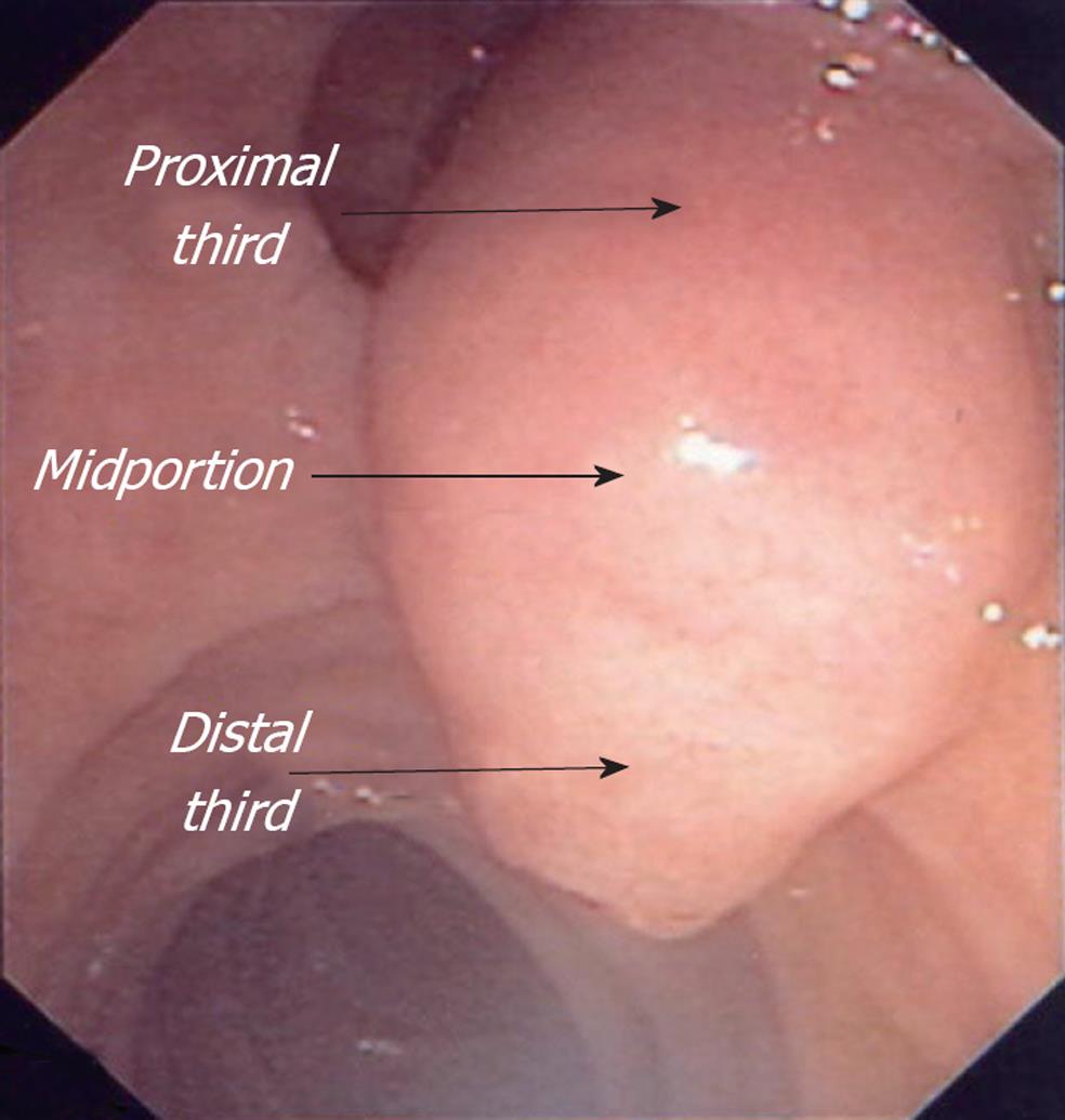
Figure 2 The starting point of the fistulotomy on the papilla.
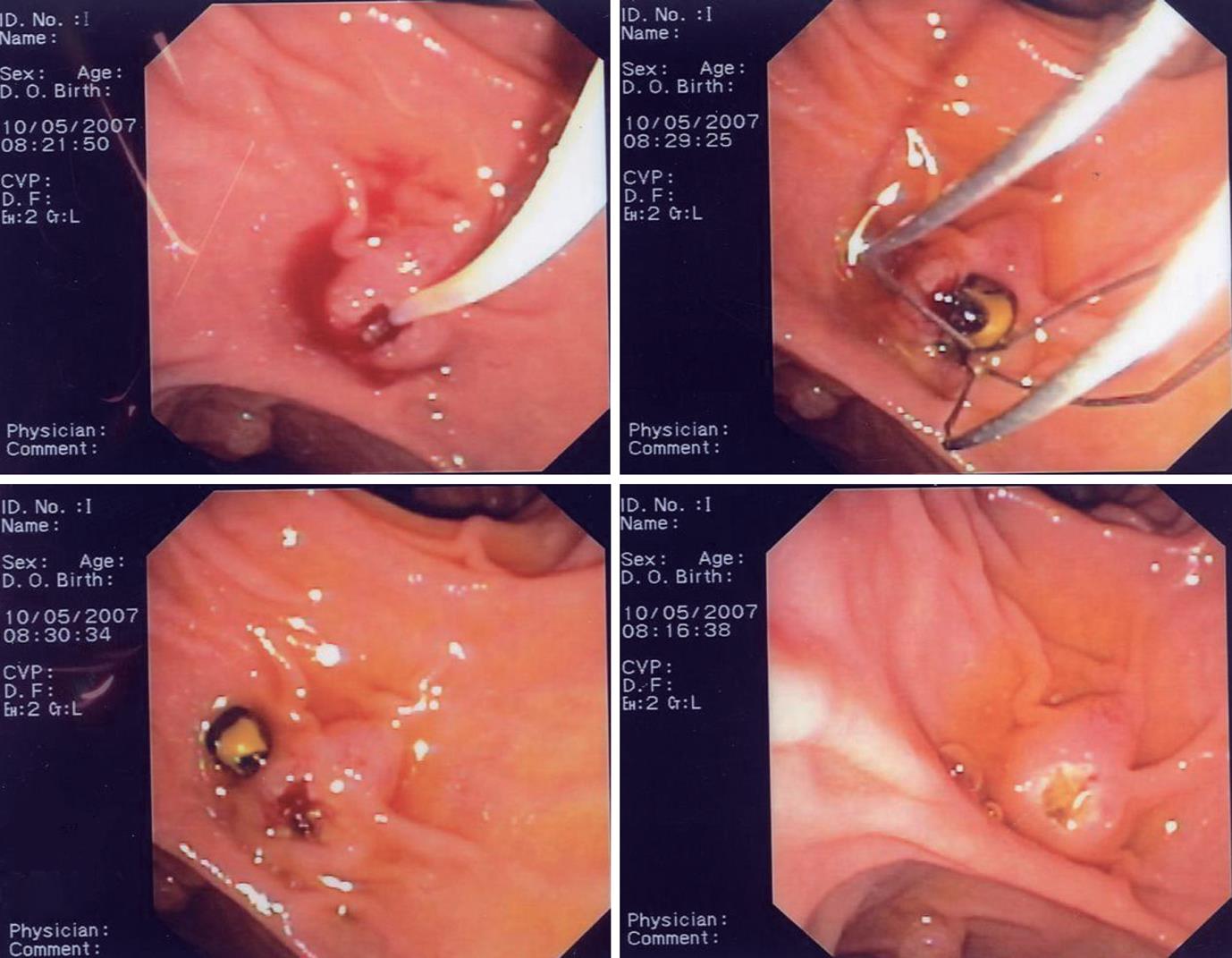
Figure 3 Needle-knife fistulotomy and placement of 10 Fr plastic stent.
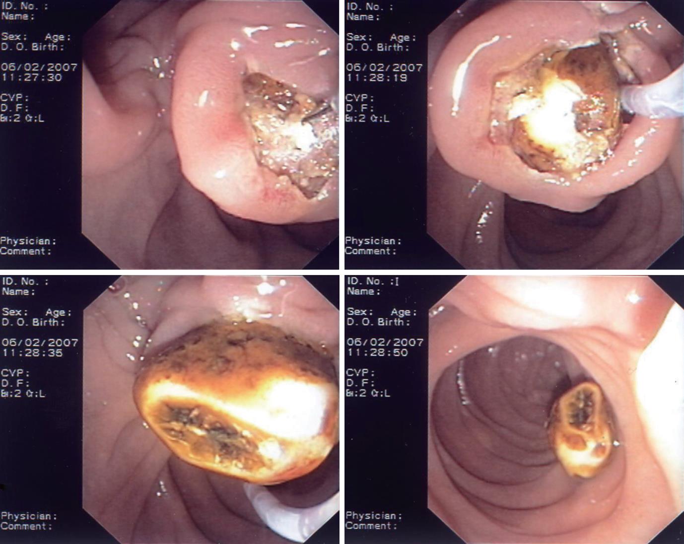
Figure 4 Needle-knife fistulotomy and removal of large stone.
Figure 5 Papilla, needle-knife fistulotomy, deep cannulation and papillotomy.
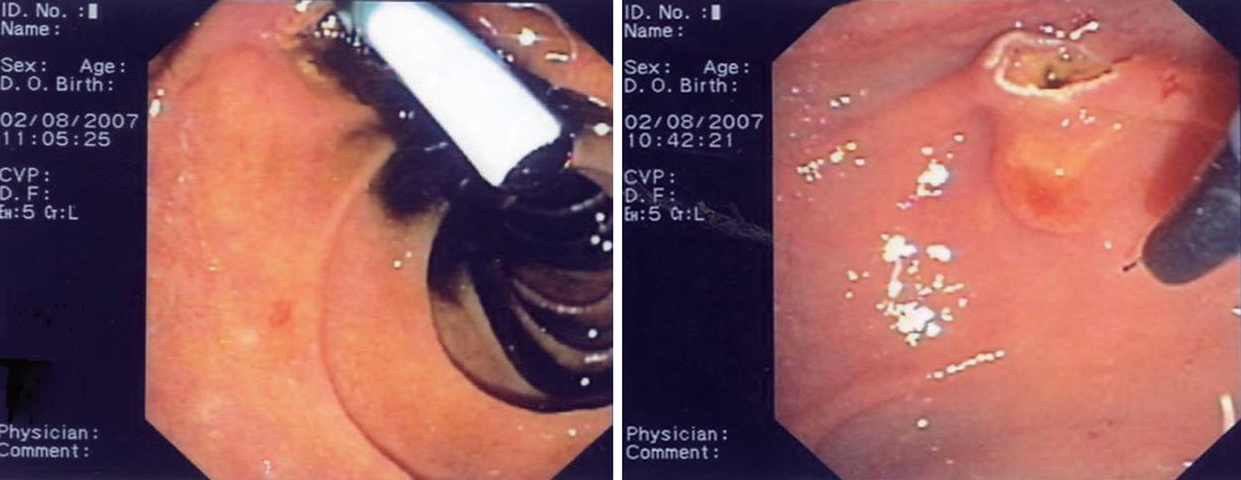
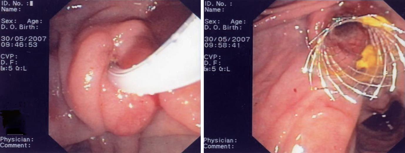
Figure 6 Needle-knife fistulotomy and placement of metallic stent.
- Citation: Ayoubi M, Sansoè G, Leone N, Castellino F. Comparison between needle-knife fistulotomy and standard cannulation in ERCP. World J Gastrointest Endosc 2012; 4(9): 398-404
- URL: https://www.wjgnet.com/1948-5190/full/v4/i9/398.htm
- DOI: https://dx.doi.org/10.4253/wjge.v4.i9.398


