Copyright
©2012 Baishideng.
World J Gastrointest Endosc. Aug 16, 2012; 4(8): 347-355
Published online Aug 16, 2012. doi: 10.4253/wjge.v4.i8.347
Published online Aug 16, 2012. doi: 10.4253/wjge.v4.i8.347
Figure 1 Eosinophilic microabscess in the esophageal superficial layer.
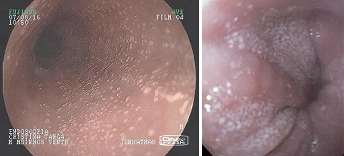
Figure 2 White specks in the mucosa of the esophagus.
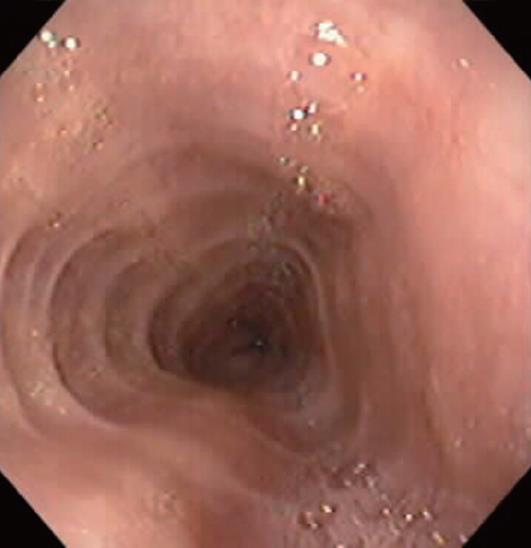
Figure 3 Rings in esophageal mucosa in eosinophilic esophagitis.
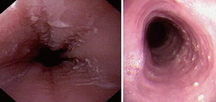
Figure 4 Linear furrowing in the esophageal mucosa.
Figure 5 White specks and linear furrowing in the esophageal mucosa.
- Citation: Ferreira CT, Goldani HA. Contribution of endoscopy in the management of eosinophilic esophagitis. World J Gastrointest Endosc 2012; 4(8): 347-355
- URL: https://www.wjgnet.com/1948-5190/full/v4/i8/347.htm
- DOI: https://dx.doi.org/10.4253/wjge.v4.i8.347


